 Newshot
Main
The presence of Homo erectus in Asia has been dated to more than 1.5 million years ago (Ma) (possibly up to 2.1 Ma if the Shangchen stone tools were made by H. erectus14), with fossil evidence coming from over 20 sites and hundreds of specimens distributed from northern China (Zhoukoudian) to Central Java (Sangiran)14,15. At a continental scale, during the Middle Pleistocene, H. erectus overlapped with Denisovans (H. longi)9,16, a hominin population now known by fossils ranging from the Altai mountains (Denisova Cave), the Tibetan Plateau and northern China to southeastern Asia (Penghu channel) and mainland southeast Asia in Laos1,7,9,17,18,19,20,21. The spatiotemporal distribution of H. erectus fossils is heterogeneous, with only a handful of late Middle to early Late Pleistocene specimens known, all located in Island Southeast Asia8,11. The latest occurrence of H. erectus is from the Late Pleistocene sites of Ngandong on Java Island, in Indonesia, dating to 117–108 thousand years ago (ka)8. By contrast, on the mainland, there are no securely dated H. erectus fossils younger than 400 ka. Specimens from the Changyang and Chaoxian caves were attributed to H. erectus, with U–Th dating of faunal remains and carbonates from these sites providing chronological ranges of 196–143 ka and 200–160 ka, respectively, for the hominin-bearing layers11,22,23. However, as these are minimum ages, the Changyang and Chaoxian H. erectus specimens could be substantially older. Moreover, molecular data are available for only a few East Asian H. erectus specimens, all dating to around 420–400 ka (ref. 13), and this limits our understanding of the evolutionary relationships of different H. erectus populations to each other and to other penecontemporary hominin populations. To address these scientific questions, we report molecular data and morphological analysis of hominin teeth from Baxian cave and Naxian cave in Guangxi Zhuang Autonomous Region, southern China (Fig. 1). These hominin teeth were recovered during recent archaeological excavations that revealed the presence of a breccia containing a combined total of over 18,000 mammalian fossils, including Elephas, Pongo and Stegodon. The two caves are located approximately 15 km apart in the same karst valley corridor, with highly similar sedimentary contexts and faunal compositions. Bayesian modelling of 43 radiometric dates provided by post-infrared infrared-stimulated luminescence dating and single-grain quartz luminescence dating of sediments, uranium series (U-series) dating of teeth, U-series dating of carbonates and coupled U-series electron spin resonance dating of teeth constrain the age of Baxian cave to 324–184 ka and the age of Naxian cave to 319–241 ka, making the two assemblages chronologically indistinguishable from each other12.
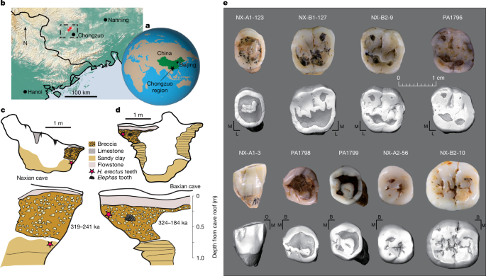
a, The location of the Chongzuo study area located in Guangxi Zhuang Autonomous Region province, southern China, marked by a black star. b, The location of Chongzuo city in relation to the dense network of karst (tan topography) that comprises this landscape. The study region has been marked by a black dashed box and the cave locations with red stars. c, Stratigraphic profile of the fossil chamber for Naxian cave showing the location of the flowstone, breccia and underlying sandy clay, and a composite stratigraphy showing the location of the H. erectus teeth and the modelled age range for the site. d, Stratigraphic profile of the fossil chamber for Baxian cave with a composite stratigraphy showing the location of the H. erectus and Elephas teeth and the modelled age range for the site. a–d were redrawn from ref. 12, Springer Nature Limited. e, Tooth images. NX-A1-123, P4; NX-B1-127, M1; NX-B2-9, M1; PA1796, M2; NX-A1-3, C1; PA1798, P3; PA1799, P4; NX-A2-56, P4; NX-B2-10, M2. M, mesial; L, lingual; O, occlusal; B, buccal.
Dental morphological analysis
The nine hominin teeth from Baxian cave (specimens with prefix PA) and Naxian cave (specimens with prefix NX) represent different tooth positions (NX-A1-123 (a fourth upper premolar, P4), NX-B1-127 and NX-B2-9 (first upper molars, M1), PA1796 (M2), NX-A1-3 (a first lower canine, C1), PA1798 (P3), NX-A2-56 and PA1799 (P4), NX-B2-10 (M2); Fig. 1 and Supplementary Note 1). All of the specimens from these two sites exhibit relatively large dimensions, falling within or close to the size distribution range of H. erectus, but also overlapping with non-erectus Asian Middle Pleistocene Homo and Neanderthals (H. neanderthalensis), and being in the higher end of the Pleistocene and Holocene modern human (H. sapiens) range of variation (Supplementary Note 1 and Supplementary Dataset 1). The two M1 teeth from Naxian cave (NX-B1-127 and NX-B2-9) are morphologically close to each other and possible antimeres of the same individual.
Among these teeth, eight show a combination of features usually found in H. erectus specimens, such as an asymmetric P4 crown24 (NX-A1-123), short upper molar crowns with a square to slightly rhomboidal occlusal outline24,25,26 (NX-B1-127 and NX-B2-9), mesial and distal marginal ridges on both lingual and buccal faces of the C1 crown26 (NX-A1-3), together with mesial and distal vertical furrows on the buccal side of C1, an asymmetric outline of the lower premolars (PA1798, PA1799 and NX-A2-56) with a slightly protruding distolingual corner of the P3 crown and a developed trigonid basin on the P4 crown24,27, a rectangular and short M2 crown (NX-B2-10) with a complex expression of the protostylid, and a distal M2 root with two canals24. Using micro-computed tomography scans to investigate the internal tooth structure, a distinctive dendrite-like enamel–dentine junction (EDJ) and shelf-like protostylid of Middle Pleistocene H. erectus from Yiyuan, Hexian and Zhoukoudian24 could be detected in the Naxian and Baxian H. erectus teeth, albeit in a more simplified form (Supplementary Figs. 3–5, 14 and 15).
This combination of features is not found in the teeth of non-erectus Chinese Middle Pleistocene Homo (such as Jinniushan, Hualongdong and Xujiayao, which probably belong to H. longi)6,9,17,24,28, nor is it present in H. neanderthalensis or H. sapiens, which each exhibit their own derived features including either a more lingually expanded or a more reduced hypocone, a simpler canine crown and EDJ morphology (even if some H. neanderthalensis canines show a developed tuberculum centrale, which is not present in the Naxian canine) or a more rounded and taller lower molar crowns6,24,28. In terms of three-dimensional enamel thickness, NX-B2-10—the only specimen suitable for 3D enamel thickness analysis—falls within the distribution range of H. erectus and differs from H. sapiens, although it approaches the boundary of the H. neanderthalensis range (Supplementary Note 1).
To quantitatively characterize the morphology of the Baxian and Naxian teeth and further confirm their taxonomic attribution, we conducted geometric morphometric analyses of the EDJ. We compared them with a broad chronogeographical sample including African and Asian H. erectus, European H. neanderthalensis, H. longi from Laos, and Pleistocene to Holocene H. sapiens from Africa and Eurasia. The results confirm the non-metric descriptions, with the EDJ of seven of the teeth being unambiguously classified as H. erectus and differing statistically from those of H. neanderthalensis and H. sapiens (Fig. 2, Extended Data Table 1 and Supplementary Note 2). The P4 specimen NX-A2-56 is statistically an outlier for all of the comparative groups (Fig. 2d). Still, the non-metric morphological features are most similar to those of PA1799, as well as earlier Chinese H. erectus specimens from Zhoukoudian and Xichuang22, characterized by a buccolingually compressed central basin with respect to the cervical outline, with the notable presence of vertical wrinkles on the mesiobuccal surface of the EDJ (Supplementary Fig. 13). The specimen also has a well-developed talonid, which is a plesiomorphic feature within Homo and therefore supports an attribution to H. erectus. Eight specimens are therefore identified as H. erectus in both metric and non-metric morphological analysis.
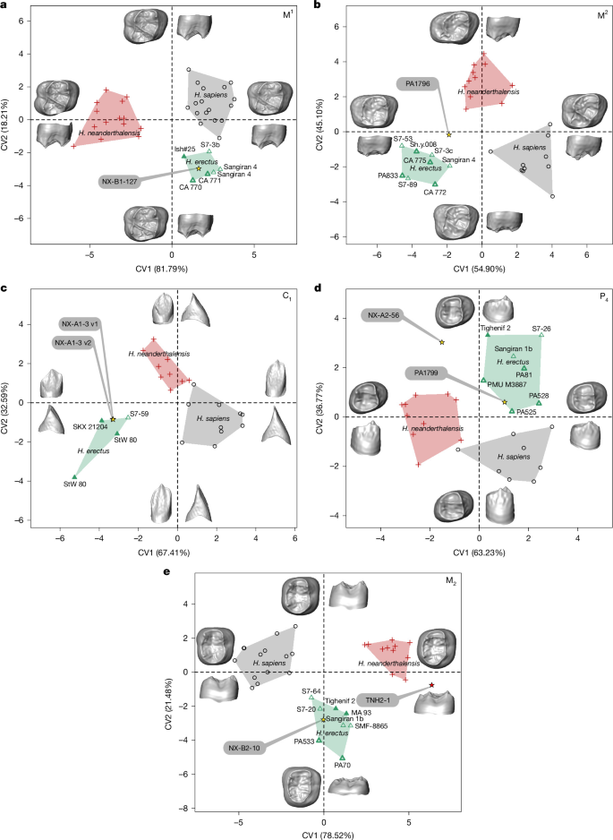
a–e, Bivariate plots of the canonical variate (CV) analyses of the EDJ shape of the Baxian and Naxian M1 (a), M2 (b), C1 (c), P4 (d) and M2 (e) compared with H. erectus s.l., H. neanderthalensis and H. sapiens. For H. erectus, the filled triangles represent African Early Pleistocene specimens, thin triangles represent Indonesian Early Pleistocene specimens and thick triangles represent Chinese Middle Pleistocene specimens. The surfaces at the end of each axis illustrate the extreme shapes along each component. Relevant statistical information is provided in Extended Data Table 1.
By contrast, the specimen PA1796 (M2) from Baxian cave displays a short EDJ with a rhomboidal outline, resulting in an intermediate shape between H. erectus and H. neanderthalensis, and being classified as compatible (P = 0.068) with the extreme limit of the H. erectus range (Fig. 2 and Extended Data Table 1). However PA1796 exhibits non-erectus non-metric features (Supplementary Notes 1 and 2). The specimen also does not fit comfortably within the morphological variation of H. neanderthalensis or H. sapiens (Fig. 2b and Extended Data Table 1). It has taller dentine horns than H. erectus, more simple trigon and talon basins (that is, without the typically wrinkled EDJ surface of H. erectus) and a more irregular EDJ outline in occlusal view; Supplementary Notes 1 and 2). It also differs from H. sapiens and H. erectus by having a more developed and lingually expanded talon.
Endogenous proteomes from southern China
To obtain molecular information on the identity of the hominin teeth, we first explored protein preservation in five faunal specimens from Naxian cave. This comparative analysis revealed that dental enamel proteomes were preserved at this site, while no endogenous dentine proteins could be detected confidently (Supplementary Notes 3 and 4 and Supplementary Dataset 2). Subsequently, we extracted dental enamel proteins from three hominin teeth from Naxian cave (P4, NX-A1-123; M1, NX-B1-127; and M1, NX-B2-9) and two hominin teeth from Baxian cave (M2, PA1796; and P4, PA1799). For both Baxian cave specimens, a small dentine sample was also taken (Methods). Combined MaxQuant, PEAKS and pFind proteomic data analysis indicates the recovery of an endogenous dental enamel proteome in all five teeth. The dental enamel proteomes are dominated by peptides deriving from amelogenins, ameloblastin and enamelin, with several additional proteins recovered from all teeth (Extended Data Table 2 and Supplementary Note 4). In comparison, peptides originating from common laboratory contaminants and human handling are low in abundance (Extended Data Fig. 1a). Unipept analysis reveals that peptide identities are to a large extent specific to Mammalia (99.5% at the class level) and Primates (98.4% at the order level), with only two peptide sequences also exactly matching UniProt entries for Fungi and three peptide sequences also exactly matching UniProt entries for Bacteria. This indicates that peptide sequence specificity is high for the dental-enamel-specific peptides identified and that the likelihood of incorrectly assigning non-primate sequences to a primate protein sequence entry are low for the recovered peptide population (Extended Data Fig. 1b,c). The recovered dental enamel peptides show short average peptide fragment lengths (Fig. 3a), advanced to saturated levels of deamidation (Fig. 3b) and elevated frequencies of other diagenetic post-translational modifications (Fig. 3c,d). N-terminal glutamines are either deamidated (between 55 and 80% depending on the specimen and analysis software used) or converted to pyro-glutamate (on average between 15 and 45%, depending on the specimen and analysis software used), with very few N-terminal glutamines not modified diagenetically (Supplementary Table 26). They also contain confident serine phosphorylation sites (localization probabilities of ≥0.95) on AMELX (Ser32), AMELY (Ser66), AMBN (Ser33, Ser41, Ser43, Ser262), ENAM (Ser54, Ser191, Ser216) and AMTN (Ser116). Serine phosphorylation is an in vivo protein modification previously observed in other hominin dental enamel proteins at the same amino acid locations29. Numerous proteins were identified in the dentine extract of PA1799, but an endogenous origin could be confirmed only for peptides deriving from COL1A1 and COL1A2, the major structural collagen present in bone and dentine (Supplementary Note 4). The proteome and peptide composition, the survival of in vivo modifications and the extent of diagenetic modifications, including extensive protein backbone hydrolysis, are all consistent with previously published Pleistocene hominid dental enamel proteomes7,29,30,31,32,33,34,35.
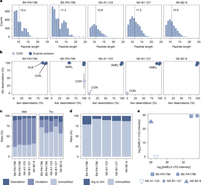
a, Peptide length distributions of dental enamel protein extracts; the average peptide length for each specimen is indicated. b, Protein-level deamidation of asparagine (Asn) and glutamine (Gln). Here, 0% indicates no deamidation and 100% indicates full deamidation. The error bars represent ±1 s.d. PA1796-ALB: n = 8 (Asn) and n = 9 (Gln); PA1796-AMEL: n = 104 (Asn) and n = 78 (Gln); PA1799-CON: n = 12 (Asn) and n = 8 (Gln). CON, contaminating proteins. c, The oxidation frequencies of methionine (Met) and tryptophan (Trp). d, The frequency of arginine (Arg) to ornithine (Orn) modification. e, Genetic sex assignment based on label-free quantification of AMELX- and AMELY-specific peptides. All panels are based on MaxQuant data analysis and, where applicable, use peptide intensities.
Four of the five teeth, PA1796, PA1799, NX-B1-127 and NX-B2-9, contain peptides uniquely matching to amelogenin-Y, a protein transcribed and translated from AMELY, a gene located on the hominin Y chromosome (Fig. 3e and Extended Data Fig. 2). These teeth therefore derive from genetically male individuals36,37,38. Conversely, for the fifth tooth, NX-A1-123, no such peptides were identified in any of the three search strategies, despite overall similar protein preservation in this tooth. We therefore identify this specimen as a genetic female. Together, this indicates that endogenous peptides were preserved in and identified from five hominin teeth, four males and one female, from Naxian and Baxian caves.
A novel H. erectus hominin lineage
Our palaeoproteomic data analysis strategy used PEAKS, pFind and MaxQuant in parallel. Sequence coverage and protein identification were highly consistent among all search strategies used (Supplementary Notes 3 and 4). PEAKS, pFind and MaxQuant sequence reconstructions agreed in all recovered amino acid positions except for one, which was subsequently excluded from the generated consensus sequences for each specimen. In total, between 566 and 1,440 (including collagen type I), or between 556 and 808 (excluding collagen type I), amino acid positions could be reconstructed (Extended Data Table 2 (tab 1)). In an Asian context, this sequence coverage is comparable to the dental enamel proteomes recovered from the Middle Pleistocene Denisovan Penghu mandible (695 positions)7, the Middle Pleistocene Denisovan from Tam Ngu Hao 2 (558 positions)6, the Denisovan Harbin dental enamel proteome (903 positions), the Middle Pleistocene H. erectus specimens from Zhoukoudian and Sunjiadong (440–599 positions)13 and the range of amino acid positions recovered from Pleistocene Pongo from East and Southeast Asia32,33, but above the number of positions reconstructed for an Early Pleistocene Gigantopithecus specimen from southern China30 and two Middle Pleistocene H. erectus specimens from Hexian13. Among the consensus sequences generated for each hominin tooth, we find no positions uniquely derived in the genus Pongo, conclusively rejecting an attribution of these teeth to the orangutan lineage (Table 1).
Protein sequence variation among Neanderthals, Denisovans and modern humans is known to be low for dental enamel proteins29,39,40. For the protein regions reconstructed here, in our reference database, the single available Denisovan reference proteome has two uniquely derived substitutions (AMBN M273V and COL1A2 R996K; Table 1). For AMBN M273V, all five individuals from Naxian and Baxian caves carry the ancestral methionine, while the single individual with coverage for COL1A2 R996K, PA1799, also has the ancestral arginine for this position. No other amino acid positions uniquely derived in Early Pleistocene hominins, Neanderthals or modern humans are covered by the data obtained from the studied fossil specimens.
Our analytical strategy revealed the presence of a single amino acid polymorphism at position 253 of ameloblastin (AMBN, UniProt:Q9NP70), where an alanine is replaced by a glycine. The substitution was independently picked up in initial PEAKS analysis for Naxian and Baxian caves, and subsequently confirmed by MaxQuant and pFind analysis (Fig. 4 and Extended Data Fig. 3). Four out of five studied hominin teeth (NX-A1-123, NX-B1-127, NX-B2-9 and PA1799) carry peptides uniquely matching the derived version of the sequence, AMBN(A253G), which probably results from a Cys to Gly transition in the codon GCC to GGC (chromosome 4: 70603880–70603882). The peptides matching this novel derived substitution do not match to known bacterial or fungal sequences. The substitution is not present in the available reference genomes or proteomes for Neanderthals (Vindija 33.19 (ref. 41), Chagyrskaya (ref. 42), Denisova 5 (ref. 43)) or the Denisovan high-coverage genome (Denisova 3)44. The ancestral alanine is also the amino acid previously reported for the dental enamel proteomes from the Penghu 1 and TNH2-1 Denisovans6,7. The substitution is absent in the great ape reference proteomes available to us45, nor is it present in the human gnomAD exome database (v.4.1, 1,461,678 individuals) or the 1000 Genomes and SGDP datasets45,46,47. The substitution was not observed in the H. erectus proteome published for D4163, a specimen dated to approximately 1.77 Ma from Dmanisi, Georgia, which contains the ancestral alanine29. However, the derived glycine was observed in six H. erectus teeth dating to approximately 0.4 Ma from East Asia. The AMBN A253G substitution therefore appears to be widespread among East Asian H. erectus13. Although the derived AMBN A253G substitution is so far exclusively observed among East Asian H. erectus specimens, this does not exclude the possibility that other specimens belonging to this population carried the ancestral amino acid variant.
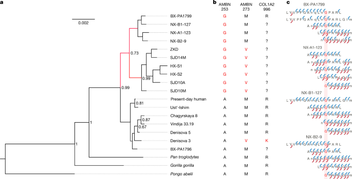
a, Bayesian phylogenetic tree using all proteins considered endogenous. Posterior probabilities lower than 0.5 are not shown. In this tree, Denisova 3 is the only included Denisovan individual, while ZKD, SJD14M, HX-S1, HX-S2, SJD10A and SJD10M refer to H. erectus specimens studied previously. b, Amino acid occupancy for reference great ape specimens, including extinct and extant hominins, for three selected dental enamel protein positions relevant for the five Naxian and Baxian hominin specimens studied proteomically in this study. c, Selected peptide-spectrum matches showing coverage of the H. erectus-derived AMBN-253G substitution observed in four of the Naxian and Baxian hominins. Peptide fragment ion series extracted from PEAKS Online v.12.
Although individual variation in protein amino acid sequences can exist between individuals, we find, among the regions of the proteins covered in our ancient data, only a single position with sequence variation among the worldwide distributed modern humans included in our database search (Table 1). In general, among extant hominids, out of the 43 amino acid positions variable among and between great ape individuals included, and in cases in which we have coverage for at least one of the four hominins studied here, only five positions contain sequence heterogeneity between the included individuals where at least four or more individuals of a single genus share variation (one position in H. sapiens, three in the genus Pan and one in the genus Pongo; Table 1). Notably, the four teeth carrying the derived AMBN A253G substitution shared with the previously reported H. erectus specimens from Zhoukoudian, Sunjiadon and Hexian differ from those six specimens, as the Naxian and Baxian hominins do not carry the derived AMBN M273V substitution (Table 1, Fig. 4 and Extended Data Fig. 3). Thus, the observation of the AMBN Gly253 substitution combined with the observation at AMBN Met273 in four hominin teeth, of at least three individuals, if the two M1s from Naxian cave represent the same individual, from two late Middle Pleistocene sites suggests that the hominin population to which they collectively belonged has not previously been sampled using molecular methods, either genetic or proteomic.
To further explore the phylogenetic placement of the Naxian and Baxian specimens, we conducted a Bayesian phylogenetic analysis. Note that, owing to the sparseness of dental enamel proteomic data, such analyses in palaeoproteomics generally have to assume that there has been no recent gene flow between the individuals and populations included in the analysis. We find that the four Naxian and Baxian individuals with the AMBN A253G substitution cluster together in a clade outside the branch of previously reported Denisovans, Neanderthals and modern humans (Fig. 4 and Extended Data Figs. 4 and 5). The topology of the clade formed by Denisovans, Neanderthals and modern humans matches the expected configuration in the main analysis, albeit with low internal node support (Fig. 4). The topology of these three hominin populations changes once additional modern human and great ape individuals are added to the phylogenetic analysis (Extended Data Figs. 4 and 5). The expected topology of this clade is challenging to obtain when using proteomic data from dental enamel proteins, as reported previously29,39,40, and it is evidenced by the low support value of this clade in our analyses (Fig. 4 and Extended Data Figs. 4–6). Nevertheless, the values supporting the nodes for the remaining branches, including the one placing the Naxian and Baxian specimens as an outgroup to the other hominins, are high in all analyses (>0.995). The Naxian and Baxian individuals are placed in a clade together with six previously reported H. erectus specimens. Those six specimens are placed together with strong support and separate from the four Naxian and Baxian individuals carrying the AMBN A253G substitution (Fig. 4). This strengthens the notion that these four Naxian and Baxian cave specimens represent a H. erectus hominin population that has not been sampled molecularly before. Furthermore, the phylogenetic analysis suggests that, although based on sparse data, this hominin population might have diverged before the divergence of the common ancestor of modern humans, Neanderthals and Denisovans. These phylogenetic observations are in accordance with the morphological analysis of these specimens, strengthening the case that these specimens belong to a previously unsampled lineage of H. erectus.
By contrast, proteomic analysis of PA1796 from Baxian cave reveals that this specimen does not contain the derived AMBN A253G amino acid substitution (Extended Data Fig. 6b). It has no uniquely derived substitutions compared to the other four teeth, nor does it share uniquely derived amino acid substitutions with the Denisovan, Neanderthal or modern human individuals present in our reference panel (Table 1). Phylogenetic analysis of this specimen places it outside of the H. erectus clade, with varying topologies with low support in relation to its placement in relation to archaic hominins (Denisovans and Neanderthals) or the clade including Neanderthals, Denisovans and modern humans (Fig. 4 and Extended Data Fig. 6a). The protein sequences reconstructed for this tooth are therefore different from the other four specimens studied proteomically, mirroring the morphological analysis that also indicates this specimen represents a taxon other than H. erectus. Only a single Denisovan genomic reference is currently available, the D3 specimen from Denisova cave, Russia, implying that our understanding of protein sequence polymorphisms for Denisovans is by definition limited. Future morphological and genomic or proteomic research on hominin teeth from the region could provide additional frameworks to allow more precise taxonomic classification of PA1796 into a non-erectus group, representing either southern Asian representatives of Denisovans (H. longi) or potentially another population or species.
Implications for East Asian H. erectus
Eight of the late Middle Pleistocene Homo teeth from the Naxian and Baxian caves exhibit metric and morphological features that lie within the range of East and Southeast Asian H. erectus and that differ from those of Denisovans (H. longi), Neanderthals (H. neanderthalensis), or Pleistocene and Holocene modern humans (H. sapiens). The palaeoproteomic evidence provides further confirmation that four of these teeth indeed fall outside of the known genetic and proteomic sequence variation of H. neanderthalensis, H. longi and H. sapiens, but are consistent with a H. erectus-derived substitution recently described for Chinese specimens dating to around 0.4 Ma (ref. 13). The recovered proteomic evidence is therefore compatible with the morphological data. Dated to between 324 and 184 ka (ref. 12), these H. erectus teeth substantially extend the known temporal range of H. erectus fossils on the Asian mainland, bringing the extent of the mainland H. erectus fossil record closer to the latest known Javanese H. erectus, dated to 117–108 ka (ref. 8). The recovery of a unique proteomic signature of mainland H. erectus from the Middle Pleistocene of East Asia furthermore suggests that specimens attributed to this species could harbour several hominin populations13, and make it possible to molecularly test whether other H. erectus fossils from the region that could potentially date to the late Middle Pleistocene, such as those from Changyang cave (Hubei province, China)11,22 and Chaoxian cave (Anhui Province, China)23, also represent specimens belonging to the same mainland H. erectus hominin population, or whether they have a proteomic signature more similar to earlier H. erectus13.
Hominins in spatiotemporal coexistence
The discovery of the Naxian and Baxian H. erectus specimens in the late Middle Pleistocene of southern East Asia enriches the hominin occupation scenario in the region. A Denisovan mandible has been described from the Penghu channel off the coast of Taiwan, dating to either 190–130 or 70–10 ka (refs. 7,48). In Laos, based on morphological analysis, a Denisovan molar has been described from Tam Ngu Hao 2, dating to between 163 and 131 ka (ref. 6). Genetic evidence on the timing of Denisovan introgression into the genomes of living humans also suggests that Denisovans were present in East and/or Southeast Asia well into the Late Pleistocene1,49. Moreover, several craniodental remains, such as Harbin, Penghu 1 and Xiahe 1, were identified molecularly as H. longi (Denisovans)7,9,17,19, and several other late Middle Pleistocene specimens probably belong to this population as well (for example, Xujiayao and Hualongdong), including also some non-erectus fossils from southern Asia (for example, Maba, Narmada, TNH2-1)6,50. It therefore seems plausible that the H. erectus population represented by the fossils from Naxian and Baxian cave on the mainland existed in relatively close proximity to Denisovan hominins.
Even though the geometric morphometric analysis of the EDJ suggest that the PA1796 M2 retains plesiomorphic aspects (mostly due to the short EDJ height), the specimen also differs from the morphology of the comparative H. erectus sample (Supplementary Note 2). The lack of a comparative sample for Denisovans (H. longi) represents a substantial bias in our ability to morphologically identify them. The non-metric morphology of PA1796 differs markedly from that of Chinese H. erectus by the lack of wrinkling on the EDJ, the absence of accessory fossae, the relatively large talon and the lingually shifted hypocone25,26 (Supplementary Notes 1 and 2). Moreover, the palaeoproteomic analysis of PA1796 shows a different signal compared to the other Baxian and Naxian specimens, as well as to the six H. erectus teeth from Zhoukoudian, Hexian and Sunjiadong13 (Fig. 4), strongly suggesting the presence of a hominin population other than H. erectus in Baxian cave. However, among the protein sequences available to us now, the PA1796 specimen does not exhibit the known proteomic variation associated with Denisovans, although it should be acknowledged that only a single Denisovan reference genome is currently available. PA1796 could therefore either represent a still-unrecognized H. erectus group that would differ in molecular and morphological aspects from the currently known H. erectus specimens, a hypothesis that we consider unlikely (Supplementary Note 2) but cannot outright reject, or belong to a non-erectus group, possibly related to H. longi. Future morphological and proteomic research on hominin teeth from the region, as well as the availability of a growing number of Denisovan genomes and proteomes, could therefore provide additional frameworks to allow a more precise classification of the PA1796 specimen.
The H. erectus specimens from Baxian and Naxian caves extend the geographical and temporal extent of mainland Asian H. erectus well into the late Middle Pleistocene. Occupying rainforest environments12, these H. erectus populations probably co-existed in close spatial and temporal proximity to southern Asian Denisovans. Mainland Southeast Asia therefore appears as a key region to study the evolution, genetics and archaeologically visible behaviours of multiple sympatric hominin species.
Methods
Metric and non-metric morphological analysis
The Baxian and Naxian hominin specimens were scanned using X-ray microtomography using the 225 kV micro-computed tomography scanner at the Institute of Vertebrate Paleontology and Paleoanthropology (IVPP, Beijing, China) according to the following parameters: 140 kV voltage, 120 µA, 0.5 angular increment one step and 360° of rotation. The final volumes were reconstructed with isotropic voxel sizes of 15.7–32.0 µm. A semi-automatic threshold-based segmentation was carried out in Avizo v.7.1 (FEI Visualization Sciences Group) and a volumetric reconstruction was then generated for each specimen. A full morphological description of each tooth can be found in Supplementary Note 1.
3D dental measurements
Dental surface models were imported in Geomagic Design v. X 64 (3D Systems, Rock Hill) to separate the crown from the tooth roots. The crown and roots were digitally isolated at the cervix using the cervix-best-fit plane as a reference51. Five variables were then measured or calculated: (1) Ve, the volume of the enamel cap (mm3); (2) SEDJ, the surface area of the EDJ (mm2); (3) Vcdp, the volume of the crown dentine and pulp (mm3); (4) 3DAET, 3D average enamel thickness (Ve/SEDJ; mm)52; (5) 3DRED, the ratio of 3D average enamel thickness to 3D average dentine thickness (Ve1/3/Vcdp1/3 × 10)53.
Geometric morphometrics
Diffeomorphic surface matching was applied to analyse the EDJ shape of the least worn teeth from Baxian and Naxian caves (methodological details are provided in Supplementary Note 2). This landmark-free, mesh-based approach was previously demonstrated to efficiently distinguish the EDJ of various hominin species6,22,27. Owing to occlusal wear, the apex of the dentine horns of some of the specimens had to be reconstructed before the analysis (Supplementary Note 2 and Supplementary Figs. 18 and 21). We first confirmed that the EDJ of the Baxian and Naxian specimens differed from that of Pongo and belonged to Homo (which was also confirmed independently by palaeoproteomics). We then compared the Baxian and Naxian EDJs to those of H. erectus s.l., H. neanderthalensis, H. longi and H. sapiens. The H. erectus sample includes dental remains from African and Asia that are regarded by some as belonging to H. ergaster and H. erectus s.s. (Supplementary Table 3). We indicated the labels of the H. erectus specimens in Fig. 2 to show the distribution of the African and Asian material in the morphospace. Canonical variate analyses were conducted and the Baxian and Naxian specimens, as well as the Denisovan molar from Tam Ngu Hao 2 (ref. 6), were projected a posteriori into the canonical variate analysis morphospace. Typicality probabilities were computed for the Naxian and Baxian teeth to provide a quantitative estimation of their most likely taxonomic attribution.
Proteomic sampling and extraction
For Naxian cave, fauna and hominin specimens were sampled using a handheld drill with a diamond-tipped drill bit, resulting in between 10 mg and 97 mg of powder for the faunal samples and up to 6 mg of hominin dental enamel. For Baxian cave, dentine was obtained using a handheld drill (36.5 and 43.2 mg of powder), while the dental enamel was sampled using an acid etch. All sampling and extraction steps were conducted in ancient molecular laboratories regularly used for ancient DNA and/or ancient protein analysis, including standard procedures to prevent contamination from occurring in the sampling and extraction environments, including wearing full body suits, hairnets, multiple layers of nitrile gloves, footies and positive air pressure.
Dental enamel extracts for Naxian cave were generated using 5% HCl, following previously published protocols31,54. Likewise, dental enamel extracts from the acid etching of the Baxian cave specimens followed previously published protocols54. The dentine samples of fauna from Naxian cave were extracted using a variant on previously published EDTA-based protocols55. For Baxian cave, the hominin dentine extractions followed a HCl-based protocol commonly used for ZooMS analysis, but with modifications to separate protein fragments by size9,56. Full protein extraction protocols are provided in Supplementary Note 3.
Proteomic data acquisition and analysis
Faunal extracts from Naxian cave were separated on the EASY-nLC 1200 (Thermo Fisher Scientific) system coupled to a Exploris 480 Orbitrap mass spectrometer (Thermo Fisher Scientific). Hominin dental enamel extracts from Naxian cave were analysed on the Vanquish Neo UPLC (Thermo Fisher Scientific) system coupled to a Exploris 480 Orbitrap mass spectrometer (Thermo Fisher Scientific). Dentine extracts from Baxian cave were first analysed on the Bruker Autoflex MaX MALDI-ToF MS instrument. Subsequent to the detection of collagen peptides, both the dentine and the dental enamel extracts from Baxian cave were analysed on an Orbitrap Fusion Lumos mass spectrometer (Thermo Fisher Scientific) coupled to an Easy n-LC 1200 HPLC system (Thermo Fisher Scientific) over a 120 min gradient. Blank extractions were included in all cases to monitor the existence of exogenous contaminants during the procedure.
Raw data files were searched separately by tissue type and cave site, using PEAKS, pFind and MaxQuant in parallel. Initial searches in PEAKS revealed the possibility of a unique substitution in AMBN, while PEAKS and pFind informed on the composition of post-translational modifications (PTMs) that should be taken into account. After initial searches, final searches using an updated protein sequence database and updated PTM information for dental enamel and the full human proteome for dentine were performed (further details are provided in Supplementary Note 3).
Protein deamidation was calculated according to a previous study for MaxQuant data57. Other PTM ratio analyses were conducted by peptide-spectrum match spectral counts or peptide counts, unless indicated differently (applying to MaxQuant, pFind and/or PEAKS). Unipept analysis was carried out using all peptide matches against endogenous, dental-enamel-specific proteins58. As described above, a project-specific database was used for data analysis that includes protein sequence variation beyond that available within UniProt. As a result, Unipept analysis will not be able to match all peptides to the same taxonomic level as the one present in the protein sequence database used. Sequence reconstruction was performed for pFind, PEAKS and MaxQuant data jointly, resulting in a single protein sequence reconstruction for each specimen. Specifically, sequences from different search engines were overlaid for each specimen to generate a single protein consensus sequence. Amino acid positions where PEAKS, pFind and MaxQuant disagreed with each other were not called, and reversed to ‘X’. Positions possibly phylogenetically informative were manually inspected, fragment ion series were checked and E values were assessed. Where necessary, positions potentially introducing invalid sequences were subsequently removed from the protein sequence reconstructions. The consensus sequences were subsequently aligned to a subset of the PaleoProPhyler database of hominid dental enamel sequences, which included four individuals from each of the genera Pongo, Gorilla and Pan, as well as three Neanderthals41,42,43, one Denisovan44 and three present-day H. sapiens from the sub-Saharan Yoruba population. We also added the dental enamel protein sequences for Homo antecessor29 and Penghu 1 (ref. 7) (a Denisovan individual) and carried out Bayesian phylogenetic analysis of the Naxian–Baxian individuals using a combination of different proteins for each tree. The combinations are indicated in each figure (Extended Data Figs. 4–6 and Supplementary Figs. 33–43). The available H. erectus protein sequences from a single specimen (D4163) from Dmanisi, Georgia, were not included in the phylogenetic analysis carried out here because the phylogenetic position of the specimen could not be resolved in the original study29.
Reporting summary
Further information on research design is available in the Nature Portfolio Reporting Summary linked to this article.