 Newshot
Main
Pokkesviricetes is a class of eukaryotic viruses with large, linear double-stranded DNA genomes within the phylum Nucleocytoviricota. The class is divided into two orders, Chitovirales (poxviruses) and Asfuvirales10. Poxviruses infect a broad diversity of animals including humans11. They include the now eradicated smallpox virus, monkeypox viruses that continue to sporadically infect humans, and molluscum contagiosum virus, causing benign tumours in immunocompromised individuals, as well as vaccinia virus, a classic model of molecular virology12,13,14. Asfuviruses infect hosts ranging from unicellular eukaryotes to animals15 and include the African swine fever virus, which causes deadly and economically important outbreaks in pigs and boars16, as well as a wider range of viruses mostly characterized in coculture with free-living amoeba15,17 or directly from the environment (for example, from plankton) using genome-resolved metagenomics6,18,19. Apart from the few viruses of global concern, as declared by the World Health Organization, the class Pokkesviricetes is of great importance for our understanding of the diversity, ecology and evolution of eukaryotic, and in particular animal, DNA viruses20,21.
The evolutionary trajectory of nucleocytoviruses leading to the emergence of animal-infecting poxviruses remains enigmatic. Animals emerged roughly 700 million years ago22 and host a variety of symbiotic unicellular eukaryotes that for the most part reside in their digestive system23,24,25, representing prime candidates for the ancestral hosts of poxviruses. Indeed, host transition from unicellular eukaryotes to animals would be greatly facilitated by a lasting period of close interactions allowing adaptation to the internal environment of the latter. Yet decades of investigations using cultivation, PCR surveys, serology and metagenomics indicated that nucleocytoviruses are mainly transient and thus rarely abundant in the digestive system of humans and other animals5,6,26. Until now, not a single group of Nucleocytoviricota was found to be commonly associated with or prevalent in the digestive system, contrasting with the broad distribution and remarkable diversity of these viruses in the environment4,6,27. With no evidence of nucleocytoviruses systematically infecting unicellular eukaryotes inside animals, early steps surrounding the emergence of poxviruses could not be explained.
Here we explored the global diversity and distribution of Pokkesviricetes by surveying tens of thousands of metagenomic assemblies from a broad range of environmental samples and animal-associated microbiomes. We discovered a third Pokkesviricetes order that we propose naming Egovirales. Egoviruses have large genomes and are predicted to form multilayered icosahedral capsids. They are the closest relatives of poxviruses and almost exclusively occur in the digestive system of humans, livestock and wild animals worldwide. One egovirus population is estimated to be present in more than 1% of the human population across continents. Highly conserved homologues of egovirus core genes were identified in genomes of Trichostomatia (ciliates), Parabasalia (Metamonada, including the pathogen Trichomonas vaginalis) and Parabasalia-associated polintons, another lineage of DNA viruses that integrate into the host genomes28, suggestive of a long history of interactions between egoviruses and these animal-associated unicellular eukaryotes. Egoviruses are among the most prevalent eukaryotic DNA viruses naturally occurring in animals. They substantially expand the evolutionary scope of Pokkesviricetes and provide critical insights into the emergence of poxviruses, pointing to anaerobic unicellular eukaryotes prevalent in the digestive system as an ancestral replicative niche that favoured host transition to animal cells.
Discovery of a third Pokkesviricetes order
DNA-dependent RNA polymerases are evolutionarily informative gene markers present in most viruses of the phylum Nucleocytoviricota, including all Pokkesviricetes10,29. We used a comprehensive hidden Markov model for the largest RNA polymerase subunit A (RNApolA)29,30 to search 85,123 metagenomic assemblies covering a wide range of environmental samples as well as thousands of samples from the digestive systems of humans, livestock and wild animals (Supplementary Table 1). This search identified 1,969,342 RNApolA genes, which we linked to main cellular and viral taxa using a custom reference database (Methods). Among the Pokkesviricetes, we identified a single poxvirus most closely related to Eptesipox occurring in the gut microbiome of a marsupial carnivore from Australia31, as well as 240 asfuviruses occurring only in the environment, predominantly in aquatic ecosystems (Supplementary Table 1). These results are in line with the current knowledge of the global niche partitioning of Pokkesviricetes and contribute, alongside previous metagenomic surveys6,19, to the great expansion of the known diversity of asfuviruses in the environment.
In addition to poxviruses and asfuviruses, we identified a clade of previously undescribed DNA viruses, related to these two viral orders, that were prevalent in the digestive system and which we provisionally name egoviruses. We characterized 12 complete, non-redundant egovirus genomes (average nucleotide identity less than 98%) from the digestive systems of humans, buffalo, goats, chicken, deer and pigs. These linear genomes are 160–360 kilobases (kb) in length and contain 55 base pair (bp) inverted terminal repeats, a feature characteristic of Pokkesviricetes32,33. The egovirus genomes encompass all eight Nucleocytoviricota hallmark genes10,29 (Supplementary Table 2), with the family B DNA polymerase (DNApolB) coding region often split into two separate open reading frames, echoing trans-splicing of another critical gene (the terminase) recently discovered in mirusviruses34. We built an hidden Markov model for the egovirus double-jelly-roll-major capsid protein (DJR-MCP) to complement the RNApolA survey and further expanded the search for egoviruses across metagenomes. Overall, we identified egovirus RNApolA and/or DJR-MCP in 864 metagenomic assemblies (detection of both markers in 286 samples), mainly from digestive systems of a wide range of animals (Supplementary Table 2). The mean coverage in these 864 egovirus-positive metagenomes was roughly 10× for the egovirus RNApolA and roughly 15× for the egovirus major capsid protein (MCP) (R2 = 0.89), reaching more than 100× in some of the human, buffalo, pig and chicken samples (Supplementary Table 2). Using these 2 marker genes, we recovered more than 200 metagenome-assembled genomes in addition to the 12 complete genomes (Supplementary Table 2 and Supplementary Information). Egovirus genomes were on average 81% complete (minimal quality score of 50%; Methods) and were up to 467 kb in size (mean of 229 kb). Phylogenetic analysis of manually curated hallmark proteins confidently identified egoviruses as a distinct main clade within the class Pokkesviricetes that is most closely related to poxviruses (Fig. 1 and Extended Data Fig. 1). On the basis of these findings, egoviruses should represent a third order within Pokkesviricetes that we propose naming Egovirales. Egovirales is the only clade within the phylum Nucleocytoviricota found to be commonly present in the digestive system.
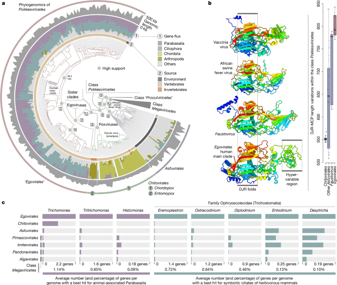
a, A maximum-likelihood phylogenomic analysis of members of Nucleocytoviricota, along with 202 Egovirales genomes with a quality score above 50%. The tree covers 1,807 genomes and was built using concatenated multiple alignments of four hallmark proteins of Nucleocytovoricota (DNApolB, RNApolA, RNApolB and transcription factor IIS; 4,110 sites) using IQ-TREE with the LG+F+R10 model. The tree was rooted with the putative class Proculviricetes and visualized using anvi’o v.9.1. Green dots in the tree highlight high supports for key nodes (approximate likelhood ratio ≥ 80 and ultrafast bootstrap approximation ≥ 95; Methods). b, Structural modelling of an Egovirales DJR-MCP (human main clade) (bottom) and comparison to the structures of DJR-MCPs from three reference Pokkesviricetes DJR-MCPs (Protein Data Bank codes from top to second from bottom: 3SAM, 6KU9 and 5J7O). The panel also shows box plots summarizing variation in protein length for the DJR-MCPs of egoviruses (complete genomes), faustoviruses, other clades of asfuviruses and Chitovirales. Centre lines in box plots show the medians; box limits indicate the 25th and 75th percentiles; whiskers extend 1.5 times the interquartile range from the 25th and 75th percentiles; all data points are visualized as individual dots. c, Gene flux signal between orders of nucleocytoviruses and eight eukaryotic genera (only the best hits are considered).
We identified 57 core genes represented in at least 10 of the 12 complete Egovirales genomes and functionally annotated most of them by sequence analysis and structural modelling (Methods and Supplementary Table 3). These core genes encompass key functionalities of nucleocytoviruses6,19,30 and include a histone doublet that we propose is involved in genome condensation into nucleosome-like structures. Histones, including histone doublets, have been previously described in some members of the Nucleocytoviricota, in particular, marseilleviruses35,36 and medusaviruses37, but not in members of Pokkesviricetes38. Egoviruses encode the main proteins involved in virion morphogenesis that are conserved across Nucleocytoviricota, including poxviruses and asfuviruses. The core components of this module are the DJR-MCP (Fig. 1), the A32-like genome packaging ATPase and the Ulp1-like capsid maturation protease. The Egovirales DJR-MCPs are unusually large (average of 806 amino acids among the complete Egovirales genomes) and the region facing the exterior of the capsid is highly diversified among egoviruses (Extended Data Fig. 2). These trends echo previous observations on the large MCPs of faustoviruses from the order Asfuvirales17. In addition, egoviruses encode homologues of the main virion proteins exclusive to asfuviruses, namely, structural polyproteins pp220 and pp62 (Supplementary Information). In African swine fever virus, both polyproteins are processed into many structural proteins that play important roles in the formation of multilayered capsids39,40. Collectively, the presence of these structural proteins strongly suggests that egoviruses form icosahedral multilayered capsids that are similar to those of Asfuvirales41, but differ from both the icosahedral capsids of most nucleocytoviruses and the brick-shaped virions of poxviruses42.
Egoviruses are globally spread in the gut
We used RNApolA and/or DJR-MCP genes of Egovirales found in metagenomic assemblies to perform a global survey of the occurrence and diversity of egoviruses (Supplementary Table 2). Both phylogenies include the same six human-specific egovirus clades (Fig. 2 and Extended Data Fig. 2). Five of these clades are specific to human gut samples, whereas the last one (clade 03) only occurred in human saliva samples. Only nine egovirus-like RNApolA genes were retrieved from environmental samples, including an aquifer, a cold seep and marine sediments (Fig. 2 and Supplementary Table 1). In the phylogenetic tree, these sequences formed four small clades, two of which (from marine sediments) were the most basal branches of Egovirales. All the remaining RNApolA sequences (more than 97% of the data) originated from the digestive system, representing the bulk of known Egovirales diversity. The DJR-MCP phylogeny recapitulated these trends, with more than 99% of the sequences, in this case, originating from the digestive system (Extended Data Fig. 3).
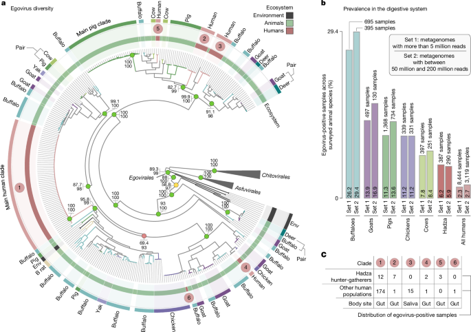
a, A maximum-likelihood phylogeny of the RNApolA of egoviruses (n = 324), using representatives of Chitovirales and Asfuvirales for rooting (1,357 sites), and the LG+F+R7 model. The layers of information summarize the ecosystem type (environment (Env) versus human versus other animals) as well as selections corresponding to egovirus clades occurring in the same animal species. The six human-specific clades are also highlighted. A similar analysis focused on the MCP of egoviruses is presented in Extended Data Fig. 3. B rat, bamboo rat. b, The prevalence for Egovirales (using signal from both the RNApolA and MCP surveys) in cohorts of metagenomic assemblies corresponding to different animal species. c, The 864 egovirus-positive samples that correspond to one of the six human-specific clades.
We surveyed 11,788 metagenomes with at least 5 million reads from 26 gut-related bioprojects (17 of them cover humans) to estimate the prevalence of egoviruses in animal species (Supplementary Table 4 and Supplementary Information). Egovirus hallmark genes were detected in the digestive system of humans of various age and geographic location (n = 193; 2.3% of egovirus-positive samples), with a notable increase of signal among hunter-gatherers from Tanzania43 (the Hadza; 6.2% of egovirus-positive samples) (Fig. 2). This enhanced signal is due to higher egovirus diversity rather than a higher prevalence of the most prevalent human clade (clade 01; just 3.1% of egovirus-positive samples). Notably, three human-specific egovirus clades were detected (almost) exclusively in the Hadza hunter-gatherers (clades 02, 04 and 05), suggesting that lifestyle could affect the diversity of human-associated egoviruses. Egoviruses were also well represented in the digestive system of pigs (n = 155; 11% of egovirus-positive samples), buffalo (n = 182; 26%), goats (n = 69; 14%), cows (n = 31; 7.8%) and chicken (n = 38; 11%). To a lesser extent, egoviruses were detected in other livestock for which fewer samples were available (deer, yak, sheep, zebu and reindeer) (Supplementary Tables 1 and 2). Similar percentages of egovirus-positive samples (for example, 2.7% for human versus 29% for buffalo) were observed when only considering metagenomes containing between 50 million and 200 million reads (n = 4,909) to minimize sequencing depth variations (Fig. 2 and Supplementary Information). Buffalo had by far the highest Egovirales prevalence among the surveyed animal species, with viruses covering a wide range of distantly related clades (Fig. 2) and prevailing in the ruminant stomach compartments (Supplementary Table 4 and Supplementary Information). Even fewer metagenomes were available for the digestive system of wild animals, but nevertheless, bamboo rats, a golden bamboo lemur, an olive baboon and a white rhinoceros were found to be extra carriers of egoviruses, indicating that primates and other wild mammals also represent a substantial reservoir of egovirus diversity.
Examination of the Egovirales dispersal across all metagenomic assemblies indicates that clades of closely related egoviruses tend to occur in the same animal species. This was most apparent for the two main clades associated with humans and pigs (Fig. 2 and Extended Data Fig. 3). We characterized the main human clade (clade 01) in 186 metagenomic assemblies from 19 studies that cover various countries from many continents, including Tanzania (the Hadza), Nepal, Fiji islanders, the USA, England, Sweden, Denmark, Germany, Italy and Spain (Supplementary Table 2). Average amino acid identity was 99.4% among the RNApolA genes and 99.9% among the MCP genes, indicating that this clade represents a cohesive viral population occurring among humans at a global scale. A complementary k-mer survey of 3 hallmark genes from a clade 01 genomic representative against assemblies of the Sequence Read Archive (SRA) detected this viral population in more than 500 metagenomes, broadening the geographic scope of its signal within the human population (Supplementary Table 4 and Methods). Overall metagenomic signal indicates that this viral population (92% of the egovirus-positive human gut samples) is present in more than 1% of the human population (including in remote rural villages and densely populated areas44,45), encompasses various stages of the human lifespan (for example, in a 3-year-old child and a pregnant woman) and remains present over time in longitudinal studies of the same individuals46 (up to nearly 1 year of sampling). The main pig clade shared similar trends, being found in 109 metagenomic assemblies from 4 studies that cover countries from many continents. Finally, the egovirus clade characteristic of human saliva was found in the USA and Fiji47,48. These results demonstrate that cohesive egovirus populations can cover expansive geographic regions while being strictly associated with a specific body site (gut or saliva) of a single animal species. In addition, we found instances of closely related egoviruses occurring in several animal species. Examples include goat–deer, pig–cow and deer–buffalo pairs (Fig. 2 and Extended Data Fig. 3). Overall, our results demonstrate the worldwide spread and strong co-occurrence of egovirus populations with one or more animal species.
Egoviruses are linked to unicellular eukaryotes
Repetitive infections over extended periods of time typically result in lateral gene transfers between viruses and their prime eukaryotic hosts, a signal detectable by means of comparative genomics6,49. To predict the host range of egoviruses, we performed a global protein-centric genomic comparison between nucleocytoviruses and a wide range of eukaryotic lineages (Methods). In line with their known host ranges, our results showed a strong gene flux signal between poxviruses and Chordata (chordopoxviruses) or Arthropoda (entomopoxviruses) (Fig. 1). By sharp contrast, the same analysis showed strong gene flux signal between egoviruses and two animal-associated groups of unicellular eukaryotes: phylum Parabasalia (43% of Egovirales–eukaryote gene flux) and order Trichostomatia (42%) (Supplementary Table 5 and Supplementary Information). This gene flux signal is effectively unique of egoviruses among the known nucleocytoviruses (Fig. 1). It is largely driven by egovirus core genes of known function (for example, DNApolB, RNA polymerase A, messenger RNA (mRNA) capping and Holliday junction resolvase) as well as egovirus core genes of unknown function (with no orthologues detected in other nucleocytoviruses), which appear to have been sporadically transferred into Parabasalia and/or Trichostomatia genomes (see Extended Data Figs. 5–7 for the corresponding phylogenetic analyses). Within Parabasalia, egoviruses were primarily linked to the genera Trichomonas and Tritrichomonas (Fig. 1 and Supplementary Table 5), which correspond to prevalent pathogens of humans and other animals. Within Trichostomatia, the gene flux signal was almost entirely constrained to the family Ophryoscolecidae (Fig. 1 and Supplementary Table 5), which is best known for its participation in digestive processes in ruminants (including buffalo)7.
Aside from Parabasalia and Trichostomatia, we did not detect any clear gene flux signal to connect egoviruses with other prevalent animal-associated unicellular eukaryotes (Supplementary Tables 5–7). The most notable case in point is Blastocystis (a stramenopile), which is widespread in the digestive system50,51,52, coexisting with egoviruses in many human, chicken and pig samples especially within the scope of our metagenomic survey (Extended Data Fig. 4 and Supplementary Table 4), and yet shows no gene flux to egoviruses. The complete lack of a gene flux signal for Blastocystis, despite a considerable niche overlap with egoviruses, quantified here with metagenomics, demonstrates that horizontal gene transfers are not random and do not occur between egoviruses and all co-occurring protists in the digestive system. Thus, although no direct evidence of infection of a specific host by egoviruses is available, the overall gene flux signal and the presence of homologues of egovirus core genes in reference Parabasalia and Trichostomatia genomes (Supplementary Table 8) appear to point to these protists as likely hosts of egoviruses. Parabasalia (Metamonada) and Trichostomatia (ciliates) belong to distinct phyla of unicellular eukaryotes53 that show commonalities in their lifestyle and cell biology. Specifically, both groups of unicellular eukaryotes have anaerobic lifestyle, inhabit the digestive tract of animals and can perform phagocytosis54,55, a mechanism well-known to be exploited by large DNA viruses to enter their host cells56,57.
Polintons contain an egovirus core gene
Polintons correspond to a main lineage of DNA viruses with a DJR-MCP that infect a wide range of eukaryotes28. Parabasalia genomes, and especially those of T. vaginalis, contain numerous endogenous polintons encompassing roughly 20 genes9. One of these genes encode KilA, a DNA-binding protein of unknown function that appears to be restricted to polintons integrated in Parabasalia genomes58. KilA is also a core gene of poxviruses and egoviruses, with a more sporadic occurrence among other nucleocytoviruses (including within the class Megaviricetes). Our phylogenetic analysis of KilA suggests an ancient evolutionary history of this gene among the nucleocytoviruses (Fig. 3). This analysis further showed that polinton KilA genes comprised two distinct clades (types I and II), with the tree topology indicating that their respective common ancestors were acquired from distinct egoviruses before the radiation of Trichomonas, Tritrichomonas and other related parabasalids from their common ancestor. In the genome of T. vaginalis strain G3 (176 Mb), the two KilA clades are represented by high copy numbers (1,116 copies of type I and 718 copies of type II) and spread across all chromosomes (Fig. 3 and Supplementary Table 9). The second species of Parabasalia with the highest copy number of the two clades is that of Trichomonas tenax. The apparent transfer of an egovirus core gene to polintons, endogenous viruses of Parabasalia, strengthens the connection between egoviruses and this clade of gut-associated anaerobic unicellular eukaryotes.
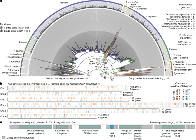
a, A phylogenetic analysis of KilA predicted proteins from nucleocytoviruses and endogenous polintons (2,207 sequences, 313 sites; model VT+F+R9), which delineates two phylogenetic clades (KilA types I and II) in the polintons. Only representatives of highly redundant Metamonada protein sequences (95% sequence identity) have been selected (Methods); the number of represented sequences is shown as ‘copy number’. b, The occurrence of the KilA genes across genomes of T. vaginalis strain G3. c, The genomic structure of polinton P1-TV (ref. 58) integrated into the genome of T. vaginalis strain G3. Chr., chromosome.
Discussion
Our global metagenomic survey of DNA viruses of class Pokkesviricetes led to the discovery of a third putative order in this class, which we provisionally name Egovirales. Egoviruses have linear genomes that can reach 467 kb in length and occur mostly in the digestive system of humans (six clades characterized), livestock (the bulk of the characterized Egovirales diversity) and wild animals, in which they prevail worldwide. Egoviruses have a highly divergent MCP (DJR-MCP). We propose that egoviruses, which encode homologues of the main virion proteins exclusive to asfuviruses, produce viral particles with an icosahedral multilayered structure resembling the virions of African swine fever virus. Egoviruses not only fill major gaps in our knowledge of the ecology and evolution of Pokkesviricetes, but also represent the only group of viruses discovered so far with large DNA genomes that is common and detected almost only in human and animal gut microbiomes. Given its evolutionary trajectory and distribution patterns, this previously overlooked main group of viruses with complex genomes is of prime interest to human and veterinary virology as well as viral evolution.
Until now, the digestive system was considered to encompass a limited diversity and abundance of eukaryotic DNA viruses5,6,26, with the most prevalent ones, such as anelloviruses59, having small, single-stranded DNA genomes of only a few kilobases. No convincing evidence of reproduction in the gut is available for viruses of eukaryotes with large DNA genomes. But, contrasting with the other Nucleocytoviricota lineages, many lines of data challenge the hypothesis of consumed water or food being the main source of egoviruses in the digestive system of humans and animals. First, egoviruses were almost never detected in environmental samples but are projected just from the metagenomic assembly signal (in the context of animal population statistics available from the United Nations) to be present in the digestive systems of roughly 180 million humans, billions of livestock animals and chickens, as well as numerous wild animals. These viruses were discovered by metagenome analysis, an approach that only targets the most abundant viral genomes and were detected by this method alone in more than 2% of the analysed human gut microbiomes and in even higher fractions of many animal microbiomes. Among animals with the highest prevalence for Egovirales, it is projected that a substantial proportion (possibly, most) of the roughly 200 million buffalo contain 1 or many egovirus species thriving in their stomach compartments. Second, the most prevalent human-specific egovirus clade alone, found in a wide range of metagenomic surveys covering several continents, is projected to be present in the gut of more than 1% of humans. In addition, longitudinal studies indicate that this clade can remain present over long periods of time in the gut of the same human individual. Thus, egoviruses are diverse, widespread and resilient viruses prevalent in the digestive system. Finally, the occurrence of one human-specific egovirus clade characteristic of the oral cavity provides the first indication that distinct egoviruses occur broadly in more animal body sites, further indicating that egoviruses are most probably not transient, but rather long-term members of the human gut and oral cavity microbiomes.
Together, comparative genomics and targeted phylogenetic analysis (for example, of the DNApolB) provide a strong prediction of virus–host interaction between egoviruses and two distantly related groups of unicellular eukaryotes (order Trichostomatia and phylum Parabasalia) that are both anaerobic, perform phagocytosis and prevail in the digestive system and oral cavity of animals24,25,54,55,60,61,62. The Egovirales–Parabasalia linkage is mostly restricted to Trichomonas and Tritrichomonas, two genera that include prominent pathogens infecting a wide range of animals and humans and can markedly affect animal immunity8. For instance, Tritrichomonas foetus is a parasite of cattle (reproductive tract) and cats (large intestine) as well as a commensal in the stomach and gut of pigs61. In addition, T. vaginalis is the most common non-viral sexually transmitted infectious agent among humans with roughly 150 million estimated cases per year63. The notable case of the apparent egovirus-to-polinton KilA gene transfer followed by the spread of polintons across the genomes of T. vaginalis and T. tenax (parasite inhabiting the human oral cavity62) further supports the long-lasting association between egoviruses and Parabasalia. The Egovirales–Trichostomatia linkage is almost entirely restricted to genera of the family Ophryoscolecidae, known to play a central role in ruminant digestive processes7,64. High prevalence of these Trichostomatia and Parabasalia genera in animals, and their phagocytosis-centric feeding lifestyle, explain the wide spread of egoviruses across animal species and suggest a long history of egovirus replication in the digestive system.
The evolution of poxviruses in animals (covering both insects and vertebrates), including the emergence of smallpox, estimated to have killed half a billion humans over the course of a century, has been extensively studied11,65,66. However, the origin of poxviruses as a group remains enigmatic due to their distant (albeit unequivocal) relationships with other nucleocytoviruses and the lack of intermediate forms. Egoviruses provide that missing link, given their phylogenetic position as the sister clade to poxviruses and their prevalence in the digestive system. Although thus far egoviruses were detected only in mammals and chickens, this signal is affected by the availability of metagenomic sequencing data among animal species, which is at present strongly biased towards humans and livestock. The diversity of egoviruses is probably substantially undersampled, especially among wild animals. The results of this work indicate that the common ancestor of poxviruses and egoviruses infected anaerobic unicellular eukaryotes capable of phagocytosis in the gut of ancestral animals before the divergence of insects and vertebrates (protostomes and deuterostomes) from their common ancestor about 700 million years ago22. Given that prominent metamonad pathogens, such as Trichomonas and Tritrichomonas, bind directly to the epithelium during infection67,68, and that pathogenic ciliates can also penetrate the animal tissue69, it is conceivable that poxviruses have evolved from an egovirus-like ancestor that gained the ability to reproduce in the animal gut epithelial cells. Our analysis of egovirus genomes suggests that the last common ancestor of Pokkesviricetes and the common ancestor of egoviruses and poxviruses had a multilayered icosahedral capsid resembling that of asfuviruses. This capsid was replaced with brick-shaped particles in the ancestors of poxviruses following their split from egoviruses and perhaps concomitantly with the invasion of animal gut epithelial cells.
The out-of-the-gut evolutionary scenario for poxviruses seems to align well with the biology of both poxviruses and eukaryotes. On the viral side, entomopoxviruses can perform an early replication phase in the midgut of insects by first infecting the epithelial cells70. There is no evidence of poxvirus replication in the gut of vertebrates. On the cellular side, phagocytosis is an important component of the immune system of animals71 serving as the entry point for various DNA viruses56,57 and is also a main feeding mechanism in both metamonads72 and ciliates73. Thus, the ability of epithelial cells to perform phagocytosis74,75 offers one possible mechanistic explanation for how a DNA virus infecting anaerobic unicellular eukaryotes in the digestive system could have successfully initiated the infection of adjacent animal cells. Altogether, this work shows that a distinct group of nucleocytoviruses is common in human and animal gut, in contrast to the previous belief, and sheds light on the origin of poxviruses.
Methods
Metagenomic database
Metagenomes (listed in Supplementary Table 1) were assembled and processed as part of the mOTUs database76,77,78. Here we provide details on the metagenomic assemblies for context. Briefly, BBMap79 (v.38.71) was used to quality control sequencing reads from all samples by removing adaptors from the reads, removing reads that mapped to quality control sequences (PhiX genome) and discarding low-quality reads (trimq = 14, maq = 20, maxns = 1 and minlength = 45). Quality-controlled reads were merged using bbmerge.sh with a minimum overlap of 16 bases, resulting in merged, unmerged paired and single reads. The reads from metagenomic samples were assembled into scaffolds using the SPAdes assembler80 (v.3.15.2) in metagenomic mode. Scaffolds were length-filtered (500 bp or more) and gene sequences were predicted using Prodigal81 (v.2.6.3) with the parameters -c -q -m -p meta.
A first metagenomic survey using RNApolA
A broad-spectrum hidden Markov model (HMM) profile for the RNApolA hallmark gene29 was searched against the predicted gene sequences from the metagenomic database using hmmsearch (HMMER82 v.3.1b2) with an E-value cutoff of 1 × 10−50. This search resulted in the identification of nearly 2 million RNApolA genes. Positive hits were then linked back to their source scaffold.
Taxonomic affiliation of RNApolA sequences
We collected amino acid sequences corresponding to a broad range of reference RNApolA genes for bacterial, archaeal, eukaryotic and giant virus lineages characterized by means of cultivation and environmental genomics30,83,84. We generated a custom diamond (v.2.1.8) database85 from this resource and summarized the taxonomic affiliation of each sequence in a complementary metadata file. We then performed a diamond blast search (v.2.1.8) of RNApolA genes identified from the metagenomic survey using this custom database and exploited the associated metadata file to assess both their taxonomic affiliation and novelty score (based on the percentage identity).
Phylogenetic signal pointing towards egoviruses
We used CD-HIT86 (v.4.8.1) and a 70% identity cutoff at the protein level to remove redundancy among the nearly 2 million RNApolA genes identified from the metagenomic survey. This step provided a database of 17,293 unique RNApolA proteins longer than 250 amino acids that encapsulate a wide range of cellular and viral diversity recovered from the metagenomic survey. Then, we used the phylogenetic workflow and visualization capabilities of anvi’o87,88 (v.8.1) using muscle89 (v.3.8.1551) and FastTree90 (v.2.1.11) to identify and remove RNApolA proteins corresponding to bacteria and plastids. Focusing on the 10,853 remaining unique RNApolA proteins, we iteratively used the same anvi’o (v.8.1) workflow of de novo alignment and tree reconstruction, but this time to identify and remove outlier sequences (isolated or long branch in the tree topology) to improve the overall quality of the tree, as commonly done in phylogenetic workflows (for example, refs. 30,91,92). A total of 13 iterations were performed, after which 8,672 unique RNApolA proteins remained. Using this curated database, we explored the phylogenetic signal of RNApolA proteins using taxonomic annotations and the novelty score for guidance. This led to the identification of a monophyletic clade corresponding to the egoviruses, which is most closely related to asfuviruses and poxviruses. We incorporated these sequences in our custom diamond (v.2.1.8) database and performed a second diamond blast on the nearly 2 million RNApolA proteins, to identify Egovirales RNApolA proteins (percentage identity more than 50%) and study their source scaffold. Data availability Item 01 provides all the RNApolA protein sequences, diamond blast databases, reference taxonomic information and fast-tree phylogenies described in this section and the previous one. This methodology recovered 12 complete Egovirales genomes.
Functional annotation of Egovirales core genes
We collected reference culture genomes and metagenome-assembled genomes (MAGs) corresponding to Asfuvirales and Chitovirales, as well as few Megaviricetes representatives from the Giant Virus Database (GVDB) collection10. We added the 12 complete Egovirales genomes into this ‘Nucleocytoviricota genomic resource’ (Data availability Item 02) and used Orthofinder93 (v.2.5.5 in diamond_ultra_sens mode) to characterize gene clusters. Gene clusters occurring in at least 10 out of the 12 Egovirales complete genomes were labelled as ‘Egovirales core genes’ (n = 57). Finally, functional annotation was performed using HHpred94 profile–profile comparisons from the HH-suite v3 package against Pfam, Protein Data Bank, Conserved Domain Database and viral protein sequences (uniprot_sprot_vir70) databases available from the MPI Bioinformatics Toolkit95. Protein structures were modelled using AlphaFold2 (ref. 96) through a local installation of ColabFold97 (v.1.5.5). In the case of the DJR-MCP of Egovirales, ColabFold97 (v.1.5.5) successfully predicted its three-dimensional structure and confirmed the presence of the DJR protein components characteristic of the phylum Nucleocytoviricota (Data availability Item 03).
Genomic completion using single copy core genes
Among the Egovirales core genes (previous section), those occurring as a single copy in at least 10 out of the 12 complete genomes were labelled as ‘Egovirales single copy core genes’ (n = 51). Notably, single copy core genes can provide relevant estimates of completion and redundancy for egovirus genomes. Briefly, we built an HMM with HMMER82 (v.3.1b2) for each Egovirales single copy core gene and created a corresponding HMM collection ready to use within anvi’o (v.8.1) (Data availability Item 04). In following sections, the completion and redundancy of egovirus genomes was predicted by counting the hits from this HMM collection, as commonly done in the field of environmental genomics and previously applied to other viral lineages (for example, refs. 30,34).
Recovery of eight Nucleocytoviricota hallmark genes
Most nucleocytoviruses contain a few core genes most central to their functioning, which are related to a virion morphogenetic module (including the MCP) and an informational module (including DNA and RNA polymerases). These genes are referred to as hallmark genes (for example, refs. 10,30). We used hmmsearch (default settings) from HMMER82 v.3.3.1 to search HMM profiles previously designed for these eight hallmark genes30, and collected matching sequences of Nucleocytoviricota from the Nucleocytoviricota genomic resource (‘Functional annotation of Egovirales core genes’ section). Given that genomes of faustoviruses from the GVDB collection10 had partial DJR-MCP genes, we replaced those with seven strains of faustoviruses from the National Center for Biotechnology Information (NCBI) GenBank collection (strains E9, LCD7, M6, S17, liban, vv10 and vv63; https://www.ncbi.nlm.nih.gov/genbank/, accessed February 2024). We also added the Variola virus isolate VARV V563 genome from the Global Ocean Eukaryotic Viral (GOEV) database30, and replaced the Megaviricetes representatives initially picked from the GVDB collection with others in the GOEV database30 on the basis of their distribution as well as the occurrence of hallmark genes. In addition to the RNApolA and DJR-MCP protein sequences already recovered, we obtained sequences for the DNApolB, the second largest DNA-dependent RNA polymerase subunits (RNApolB), the transcription factor IIS, the D5-like primase (Primase), the poxvirus late transcription factor and the packaging ATPase. The DNApolB gene, often present on two separate open reading frames in egoviruses, required manual curation through BLASTp alignments (BLAST98 v.2.10.1) and phylogenetic reconstructions, as described in ref. 30. Briefly, when a single egovirus genome had two sequences for this gene, their positions were inspected in a maximum-likelihood single-protein phylogenetic tree, and their sequences were aligned with BLASTp to a close reference sequence and to each other. The two DNApolB sequences for a same egovirus systematically corresponded to a clear split: they were fused and accordingly labelled for further notice. The same protocol was performed for the RNApolA and RNApolB protein sequences, occasionally split in egoviruses. Finally, we created a database of 77 Nucleocytoviricota genomes containing at least 7 out of the 8 hallmark genes, for downstream single-protein phylogenetic analyses (Data availability Item 05).
Alignments, trimming and single-protein phylogenetic analyses
From the genomic database created in the previous section (section ’Recovery of eight Nucleocytoviricota hallmark genes’), protein sequences corresponding to the same hallmark genes were aligned with MAFFT99 v.7.464 using the L-INS-i algorithm and default parameters. Sites with more than 70% gaps were systematically trimmed using goalign v.0.3.5 (https://www.github.com/evolbioinfo/goalign). The maximum-likelihood phylogenetic reconstructions were performed using IQ-TREE100 v.1.6.2, with the ModelFinder101 option to determine the best-fitting model. Supports were computed from 1,000 replicates for the Shimodaira–Hasegawa-like approximate likelihood ratio (aLRT)102 and ultrafast bootstrap approximation (UFBoot)103. Supports were deemed good when Shimodaira–Hasegawa-like aLRT 80% or greater and UFBoot 95% or greater, as per the IQ-TREE manual and moderate when only one of the two conditions was met. The eight phylogenetic trees (Data availability Item 06, including protein sequences and trees) were visualized and rooted between the Megaviricetes and Pokkesviricetes classes using anvi’o (v.9.1).
A second metagenomic survey using the DJR-MCP
We collected 20 DJR-MCP sequences from Egovirales source scaffolds (section ‘Phylogenetic signal pointing towards egoviruses’), which include the complete genomes. We aligned them using MAFFT v.7.464 (L-INS-I algorithm; default settings) and generated an HMM profile dedicated to the Egovirales DJR-MCP (Data availability Item 04) using hmmbuild (HMMER82 v.3.3.2). This HMM profile was then searched against all predicted gene sequences from the metagenomic database, using hmmsearch (HMMER82 v.3.3.1) and an E-value cutoff of 1 × 10⁻¹⁰. Positive hits (based on both RNApolA and DJR-MCP) were then linked back to their source scaffold, expanding the scope of Egovirales-positive metagenomic samples (Data availability Item 07).
A database of 224 egovirus environmental genomes
The mOTUs database contains metagenomic bins generated using Metabat2 (ref. 104) (v.2.15) with differential coverage information, as described in refs. 76,77. Here we analysed all scaffolds of mOTUs containing signal for the RNApolA gene and/or DJR-MCP gene of Egovirales, along with other scaffolds occurring in their associated bins. From the 57 Egovirales core genes, we selected 42 that were highly specific to Egovirales compared with Asfuvirales, Chitovirales and Megaviricetes (that is, those genes often occur in Egovirales but rarely in other nucleocytoviruses), and created a collection of 42 HMMs extracted from the collection of 57 core genes (Data availability Item 04). Scaffolds lacking signal for any of those 42 HMMs were removed to only work on high-quality viral MAGs. After this removal step, we predicted the completion and redundancy of Egovirales MAGs using the HMM collection of Egovirales single copy core genes. Finally, we removed MAGs with a quality score (completion minus redundancy) below 50%. We processed the final genomic database (Data availability Item 08, which contains 224 Egovirales environmental genomes) with anvi’o (v.8.1) to predict gene sequences (with Prodigal81 v.2.6.3) and functionally annotated hallmark genes using specific HMMs. The Egovirales proteins were subsequently clustered into orthogroups using OrthoFinder (v.2.5.5, default settings), and orthogroup assignments were mapped to those from the initial functional annotation (section ‘Functional annotation of Egovirales core genes’). Finally, we used FASTANI105 (v.1.34) within anvi’o (v.8.1) to create a non-redundant database (less than 95% average nucleotide identity) of 116 Egovirales environmental genomes, 6 of which are human specific (Data availability Item 08).
A phylogeny of Nucleocytoviricota
We performed a multi-protein phylogenetic analysis of nucleocytoviruses on the basis of the concatenation of DNApolB (sequences of 100 amino acids or more), RNApolA (sequences of 200 amino acids or more), RNApolB (sequences of 200 amino acids or more) and transcription factor IIS protein sequences (sequences of 25 amino acids or more), as previously done in ref. 30. For Megaviricetes, we used manually curated hallmark genes from the GOEV database30 (excluding Medusavirus). For the Pokkesviricetes, we used the same resource as described in the section ‘Recovery of eight Nucleocytoviricota hallmark genes’ (genomes from the GVDB collection10 supplemented with faustoviruses from the NCBI GenBank collection and Variola virus from GOEV; Data availability Item 05) and the 224 Egovirales environmental genomes (Data availability Item 08). This phylogenetic analysis followed the protocol described in the previous section, except that the FFT-NS-i algorithm of MAFFT v.7.464 was used for the alignment of each individual protein dataset, before they were trimmed (sites with 70% or greater gaps, using goalign v.0.3.5) and concatenated. Supports were computed from 1,000 replicates for the Shimodaira–Hasegawa-like aLRT102 and UFBoot103. The phylogenetic tree (Data availability Item 06) was visualized and rooted using anvi’o (v.9.1).
RNApolA and MCP Nucleocytoviricota phylogenies
To go beyond the scope of environmental genomes (previous section), we performed global phylogenies containing all RNApolA (sequences more than 600 amino acids) and DJR-MCP protein sequences (sequences more than 600 amino acids) of egoviruses extracted from the metagenomic survey (Data availability Item 07), using genomes of asfuviruses and chitoviruses from GVDB as the outgroup. The phylogenetic trees were inferred using the same approach as for the single-protein phylogenies: the sequences were aligned using the L-INS-i algorithm of MAFFT v.7.464 (default settings), sites with 70% or more gaps were trimmed using goalign v.0.3.5 and the trees reconstructed using IQ-TREE v.1.6.2 and the ModelFinder option. Supports were computed from 1,000 replicates for the Shimodaira–Hasegawa-like aLRT102 and UFBoot103. The phylogenetic tree (Data availability Item 06) was visualized and rooted using anvi’o (v.9.1).
Metagenomic signal for egoviruses and co-occurring eukaryotes
We generated a non-redundant database (average nucleotide identity less than 95%) of egovirus MCP genes, egovirus RNApolA genes and eukaryotic RNApolA genes characterized from the Egovirales-positive samples using CD-HIT86 (v.4.8.1) (Data availability Item 09). We performed read recruitment of all the Egovirales-positive metagenomic samples against the database. For that, quality-controlled metagenomic reads were mapped (Bowtie2, ref. 106, v.2.4.116) against the database and the ‘anvi-profile-blitz’87,88 (anvi’o v.8.1) routine used alignments to generate mean coverage values. Taxonomic information was included using the diamond blast (v.2.1.8) for RNApolA genes (section ‘Taxonomic affiliation of RNApolA sequences’) and the phylogenetic tree for MCP genes.
k-mer survey of the most prevalent human-related egovirus
A global k-mer survey (k = 31) of the MCP, DNApolB and RNApolA single copy core genes of a genomic representative of the main human clade (MAG: ZEEV15_1_SAMEA3664761_BIN_00000011) was performed against the SRA data (at the assembly level) using the online tool Logan-Search107, with a minimum k-mer coverage of 50% (default Logan-Search cutoff to minimize false positives). This search provided 524 candidate-positive samples, comprising 270 with 3 markers, 95 with 2 and 159 with 1. Because the tool only allows screening of nucleotide sequences smaller than 1 kb, we only analysed the first 1 kb of these three genes.
Protein sequence comparisons between viruses and cells
Egovirales proteins (from the 224 environmental genomes), together with Pokkesviricetes (Asfuvirales and Chitovirales) from the GVDB collection and Megaviricetes from the GOEV collection, were used as queries for similarity searches against UniProt-TrEMBL using MMseqs2 (ref. 108) (v.17-b804f) with default settings. Hits not clearly assigned to cellular organisms were removed on the basis of taxonomic annotations (Supplementary Table 5). For each query, only the best hit with 30% or more identity and a minimum bit score of 100 was retained. Given the enrichment in Metamonada and Trichostomatia (Ciliophora) among hits for Egovirales proteins, the same queries were searched against further metamonad and ciliophoran genomes retrieved from NCBI Genome and the Genome Warehouse (list in Supplementary Table 8; Data availability Item 10 provides the corresponding proteomes) using identical parameters. Bit score values were used to compare the hits across the independent runs, and only the highest-scoring hit per query was retained. For functional overview, Egovirales proteins and their corresponding best hits in metamonads and Ciliophora were queried for searches against Pfam-A.full (release 38.0) using MMseqs2 (v.17-b804f; default settings), retaining only the best hit per query. To further assess the specificity of the detected similarities, we performed reciprocal similarity searches using all metamonad and ciliophoran proteins that matched an Egovirales protein (30% or greater sequence identity and bit score 100 or greater) as queries, irrespective of whether they represented the best hit for the original viral query. These proteins were searched against the Egovirales, Pokkesviricetes (Asfuvirales and Chitovirales) and Megaviricetes protein datasets using MMseqs2 (v.17-b804f) with default settings (Supplementary Table 8). They were also searched against UniProt-TrEMBL to identify proteins showing stronger similarity to homologues outside Nucleocytoviricota. When metamonad proteins were used as queries, UniProt-TrEMBL hits assigned to Metamonada were excluded to avoid self-matches to closely related strains represented in the resource. For each query, only the best hit within each target dataset was retained.
Egovirus core genes found in Parabasalia and Trichostomatia
Our protein sequence comparisons identified a gene flux signal connecting egoviruses and two clades of eukaryotes: Parabasalia and Trichostomatia. Most of this signal was due to egovirus core genes occurring in the eukaryotic genomes. To clarify the directionality of this gene flux, we performed phylogenetic analyses for four egovirus core genes of known function (DNA-dependent RNA polymerase largest subunit (RNApolA), DNApolB, mRNA capping enzyme and Holliday junction resolvase) and two egovirus core genes of unknown function (OG0000040 and OG0000089). HMM profiles for the mRNA capping enzyme (PF03919.22) and Holliday junction resolvase (PF04848) were retrieved from the InterPro database (https://ebi.ac.uk/interpro/), whereas new HMM profiles were constructed for OG0000040 and OG0000089 (those are core genes of Egovirales lacking any annotation) using hmmbuild (HMMER82 v.3.3.1) from Egovirales sequences aligned with MAFFT v.7.464 (FFT-NS-2 algorithm, default settings) (Data availability Item 04). Homologous sequences were identified using hmmsearch (HMMER82 v.3.3.1) with the profiles’ gathering threshold score cutoffs for the InterPro HMM profiles (default minimum bit score of 22 for Holliday Junction and 26.5 for mRNA capping) and default E-value inclusion threshold for the two others (E-value below 0.01), and collected from Pokkesviricetes, Megaviricetes and the set of extra Metamonada and Ciliophora (previous section), as well as from the TCS database (derived from EukProt v.3 and representing the diversity of eukaryotes109). To reduce redundancy for the mRNA capping enzyme and Holliday junction resolvase, sequences were clustered with CD-HIT (v.4.8.1) with dataset-specific thresholds (80% or more and 95% identity for TCS sequences in the mRNA capping enzyme and Holliday junction resolvase datasets, respectively; 90% or more identity for GOEV sequences in both gene families). For DNApolB and RNAPa, HMM profiles and cellular and/or viral homologues were obtained from ref. 30, and homologues from metamonads, Ciliophora and egoviruses identified using hmmsearch (HMMER v.3.1) were directly incorporated into these datasets. For the DNApolB, RNApolA and mRNA capping, sequences shorter than 100, 200 and 100 amino acids, respectively, were removed. In the case of DNApolB, we also removed sequences longer than 2,000 amino acids. Sequences were aligned using MAFFT v.7.464 (FFT-NS-2 algorithm, default settings), sites with 70% or larger gaps were trimmed using goalign v.0.3.5 and phylogenetic trees (Data availability Item 06) were reconstructed using IQ-TREE v.1.6.2 with the ModelFinder option. The alignment, trimming and tree inference process was iterated for some of the trees to identify and remove outlier sequences (isolated or long branch in the tree topology) before final inference to improve the resolution and stability (Supplementary Information and Data availability Item 06).
KilA linking egoviruses to metamonad polintons
The KilA gene, which is associated with polinton-like elements in metamonads, especially parabasalids, was analysed separately due to its distinct evolutionary context. An HMM profile (PF04383.19) was retrieved from InterPro (https://ebi.ac.uk/interpro/) and used to identify homologous sequences across Pokkesviricetes (including egoviruses), Megaviricetes and Metamonada using hmmsearch (HMMER v.3.3.1, profile’s gathering threshold score cutoff). Considering the specific enrichment of polinton-like elements in Metamonada, extra genomes were included: T. vaginalis strains ATCC 30188 (GCA_002891335.1) and ATCC 50138 (GCA_052324655.1), T. tenax isolate Hs4:NIH (GCA_047301845.1) and strains NIH4 ATCC 30207 (GCA_900231805.1), Trichomonas gallinae isolate XT1081-07 (GCA_008369845.1), Trichomonas sp. genotypes 1c (GCA_047372395.1) and 2a (GCA_047372375.1), and Pentatrichomonas hominis isolate Hs-3:NIH (GCA_047301545.1). To limit redundancy, sequences from GOEV, GVDB and Metamonada genomes were clustered independently using CD-HIT (v.4.8.1; 95% or more identity threshold), and only the representative sequence of each cluster was retained (Data availability Item 11). To reduce the volume of eukaryotic sequences, all the sequences were used as queries in a similarity search against Pfam-A.full (release 38.0) using MMseqs2 (v.17-b804f; default settings); eukaryotic sequences without a best hit to KilA-N were removed. Remaining sequences were aligned using MAFFT v.7.464 (FFT-NS-2 algorithm; default settings), sites with 70% or greater gaps were trimmed using goalign v.0.3.5 and the phylogenetic trees were inferred using IQ-TREE v.1.6.2 with the ModelFinder option. Outlier sequences (isolated or long branch in the tree topology) were removed from the sequence dataset before a new iteration of de novo alignment, trimming and tree reconstruction was performed. A total of eight iterations (in addition to the initial tree) were completed (Supplementary Information and Data availability Item 06).
Genomic synteny
Putative genes for the genome of T. vaginalis strain G3 (GenBank reference GCA-026262505.1) were retrieved, including their functional annotation and predicted positions of polinton-like regions110. Genomic positions of Egovirales-like hits as well as KilA genes of type I and type II were mapped onto it. Finally, MCP genes matching that of Polinton-1_TV, a well annotated polintovirus from the same T. vaginalis strain G3 (ref. 28) collected from the REPBASE database (https://girinst.org/repbase/), were identified using MMSeqs2 (v.17-b804f) and default settings.
Reporting summary
Further information on research design is available in the Nature Portfolio Reporting Summary linked to this article.