 Newshot
Main
Amyotrophic lateral sclerosis (ALS) is a rapidly progressive and fatal neurodegenerative condition. Recently, antisense oligonucleotides (ASOs) targeting SOD1 and FUS have shown early promise in clinical trials9,10, but these approaches only benefit patients with SOD1 and FUS mutations—less than 3% of patients with ALS—leaving an urgent need for therapies for the remaining roughly 97% of patients with ALS. A common feature of all non-FUS and non-SOD1 ALS is the aggregation of TAR DNA binding protein-43 (TDP-43) in neurons1,2, a finding also observed in roughly 50% of both frontotemporal dementia (FTD) and Alzheimer’s disease2,11. However, no therapies that directly target TDP-43 are available, leaving the underlying nuclear loss of function (LoF) unaddressed. TDP-43 is an RNA-binding protein that is normally located in the nucleus and has crucial roles in the regulation of RNA splicing12,13. Cytoplasmic mislocalization of TDP-43 induces a nuclear LoF resulting in cryptic exon inclusion, cryptic alternative polyadenylation and/or exon skipping in some mature messenger RNA (mRNA) transcripts3. These events occur in hundreds of transcripts, including genes with crucial neuronal functions, such as STMN2, UNC13A and KCNQ2 (refs. 4,5,6,7,8). Whereas the impact of STMN2 mis-splicing on axonal regeneration and KCNQ2 exon skipping on intrinsic excitability has been studied, the direct impact of TDP-43 LoF on synaptic function has not been addressed.
Genome-wide association studies (GWAS) have shown that single-nucleotide polymorphisms (SNPs) located within and proximal to a cryptic exon in UNC13A increase the risk of developing ALS and accelerate disease progression14,15,16. Under normal physiological conditions, these risk SNPs do not induce inclusion of the UNC13A cryptic exon, but after TDP-43 LoF, risk SNPs exacerbate cryptic exon inclusion leading to increased loss of functional RNA transcripts and protein6,7. UNC13A is a presynaptic protein that is required at the active zone to properly dock and prime synaptic vesicles for fusion with the plasma membrane17. Genetic deletion of Unc13a in mice leads to a roughly 90% reduction in spontaneous and evoked glutamatergic excitatory synaptic transmission18,19,20. Although the UNC13A genetic association strongly implicates this event in ALS pathogenesis, the contribution of TDP-43 LoF to synaptic dysfunction and the role of UNC13A cryptic splicing—particularly in the context of many mis-splicing events in other synaptic transcripts—is unaddressed. Resolving these outstanding questions is crucial for establishing whether correcting UNC13A splicing represents a viable therapeutic approach for restoring normal neuronal function.
Here we report that TDP-43 loss in mature human induced pluripotent stem (iPS) cell-derived neurons induces loss of presynaptic UNC13A and a notable reduction in AMPA (α-amino-3-hydroxy-5-methyl-4-isoxazole propionic acid) receptor-mediated synaptic transmission, leading to a loss of coordinated network activity. Despite the widespread roles of TDP-43 in RNA regulation, isolated genomic deletion of the UNC13A cryptic exon rescues these deficits, demonstrating that this single event is a key driver of synapse dysfunction in ALS. To this end, we developed splice-switching ASOs that can rescue UNC13A mRNA splicing, UNC13A protein levels and UNC13A presynaptic localization, and restore both spontaneous and evoked synaptic transmission. Furthermore, we demonstrate that ASO treatment rescues UNC13A cryptic exon inclusion in a humanized mouse model that recreates this human-specific mis-splicing event. Thus, ASOs targeting the UNC13A cryptic exon represent an innovative therapeutic strategy for treating ALS and FTD, and their use for treating sporadic ALS is supported by genetic evidence.
Loss of TDP-43 depletes synaptic UNC13A
TDP-43 plays important roles in development and therefore, to avoid interfering with neuronal maturation by depleting TDP-43 from the iPS cell stage, we used CRISPR–Cas12 gene editing to generate a cell line in which both endogenous copies of TDP-43 harbour a HaloTag to allow for proteolysis targeting chimera (PROTAC)-mediated21 knockdown of TDP-43 in mature neurons (Fig. 1a and Extended Data Fig. 1a–d). TDP-43 is degraded by HaloPROTAC in a dose-dependent manner and in a process blocked by proteasome inhibition or the NEDD8 E1 inhibitor (Extended Data Fig. 1e–g). HaloTag-TDP-43 iPS cells were differentiated into mature excitatory cortical-like neurons (HaloTDP iNeurons) that expressed the layer V excitatory cortical marker CTIP2 and lacked expression of the inhibitory marker NKX2.1 or the neuronal progenitor marker PAX6 (Extended Data Fig. 1h). TDP-43 was also found to be correctly localized to the nucleus (Fig. 1b). Tagging of TDP-43 can alter its function and, whereas the HaloTag partially affected TDP-43 function (Extended Data Fig. 1i), UNC13A cryptic splicing and mRNA downregulation were robustly detected only on HaloPROTAC knockdown of TDP-43 (Fig. 1c,d and Extended Data Fig. 2a), similar to CRISPR interference (CRISPRi)-mediated knockdown (Extended Data Fig. 2b–f). Loss of UNC13A mRNA led to a time-dependent decrease in UNC13A protein whereby less than 5% of protein remained after 10 days of HaloPROTAC treatment (Fig. 1e,f). Immunofluorescence staining revealed a loss of synaptic UNC13A staining at synapsin-positive puncta (Fig. 1g,h). HaloPROTAC-mediated knockdown of TDP-43 in mature neurons did not lead to alterations in the RNA levels of N-methyl-d-aspartate and AMPA receptor subunits as assessed by reverse transcription–quantitative PCR (RT–qPCR) (Extended Data Fig. 2g).
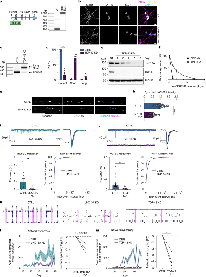
a, Schematic of HaloTag insertion at the start of TARDBP exon 2 and genomic PCR (n = 1) confirming editing. b, Immunofluorescence showing nuclear TDP-43 localization in untreated (NT) HaloTDP iNeurons and loss following HaloPROTAC treatment (n = 3). c, RT–PCR analysis of UNC13A splicing following TDP-43 knockdown. d, Quantification of c; n = 12 biological replicates from 3 experiments. e, Western blot of HaloTDP iNeurons treated with 30 nM HaloPROTAC-E for the indicated times. f, Quantification of e; n = 2 biological replicates. g, Synapsin and UNC13A immunofluorescence in 4-week-old HaloTDP iNeurons cultured on rat astrocytes after 2 weeks of TDP-43 depletion. h, UNC13A intensity at synapsin-positive terminals (control n = 116; TDP-43 knockdown n = 115 synapses; 3 experiments). i, mEPSCs from control (n = 12) and UNC13A-knockdown (n = 13) iNeurons (3 experiments), with representative and mean traces, frequency and inter-event interval cumulative distributions. j, mEPSCs from control (n = 20) and TDP-43-knockdown (n = 22) HaloTDP iNeurons (3 experiments), with representative and mean traces, frequency and inter-event interval cumulative distributions. k, Representative MEA raster plots (60 s) for control (DIV38) and UNC13A-(DIV32) and TDP-43-(DIV38) knockdown; electrode bursts, blue; network bursts, pink. l,m, Network synchrony over time in control versus UNC13A- (l) or TDP-43- (m) knockdown iNeurons; knockdown values normalized to control (n = 18 wells, 3 experiments). Symbols indicate independent experiments. In l,m, lines connect parallel differentiations recorded on the same MEA plate. Statistics: two-sided Student’s t-test (d); Gaussian linear mixed-effects model (LMM) with two-sided Satterthwaite-corrected t-tests (h–j); two-sided ratio paired t-tests (l,m). P < 0.05; P < 0.01; P < 0.001; ****P < 0.0001. Uncropped gels are shown in Supplementary Fig. 1. Scale bars, 10 µm. In all figures, error bars represent mean ± s.e.m. CTRL, control; DIV, days in vitro; FC, fold change; KD, knockdown; NS, not significant; NT, no treatment; PSI, percentage spliced in.
Loss of TDP-43 impairs synapse function
Previous investigations of the physiological functions of UNC13A have been mainly conducted using primary rodent neurons with limited studies in human neurons18,19,20,22. We therefore sought to assess the impact of UNC13A loss in human iPS cell-derived neurons (Extended Data Fig. 2h). Whole-cell patch clamp recordings showed that depletion of UNC13A through CRISPRi impaired synaptic transmission, as evidenced by a reduction in the frequency of miniature excitatory postsynaptic currents (mEPSCs) (Fig. 1i), consistent with a presynaptic deficit and in line with previously published work19,20. HaloPROTAC-mediated TDP-43 depletion also reduced mEPSC frequency (Fig. 1j), mirroring the presynaptic deficit observed on UNC13A loss. Treatment with 6-cyano-7-nitroquinoxaline-2,3-dione (CNQX) blocked mEPSCs, confirming that synaptic events were mediated primarily by means of fast AMPA receptor-mediated glutamatergic transmission (Extended Data Fig. 3a). Whereas UNC13A depletion had no effect on mEPSC amplitude, consistent with a purely presynaptic deficit (Extended Data Fig. 3b), TDP-43 depletion led to a modest reduction in amplitude (Extended Data Fig. 3c), suggesting extra postsynaptic deficits independent of UNC13A depletion. Passive membrane properties remained comparable between control and UNC13A–TDP-43 depleted neurons (Extended Data Fig. 3d,e).
To investigate the effect of UNC13A and TDP-43 loss on network activity, we carried out multi-electrode array (MEA) recordings. We found that either UNC13A or TDP-43 depletion induced a disordered and asynchronous pattern of network activity, measured by a reduction in the normalized electrode cross-correlations (Fig. 1k–m). This result indicates that UNC13A–TDP-43 depleted neurons fail to support network-level activity. The number of active electrodes remained consistent (Extended Data Fig. 3g) and phase-contrast images showed comparable cell coverage between conditions (Extended Data Fig. 3h), ruling out issues with electrode coverage as a confounding factor. CNQX treatment of control neurons confirmed network synchrony was highly dependent on fast AMPA receptor-mediated synaptic transmission (Extended Data Fig. 3f). Whereas extra MEA metrics were also affected by both TDP-43 and UNC13A loss, the network synchrony deficit was the largest and most consistently observed change (Extended Data Fig. 3g). Thus, we show that TDP-43 depletion substantially impairs fast glutamatergic synaptic transmission and synchronized network activity in human neurons, mirroring a comparable loss of synaptic function and network activity induced by direct UNC13A loss.
Genomic deletion of UNC13A cryptic exon
Knockdown of TDP-43 leads to cryptic exon inclusion within UNC13A intron 20 along with an independent downstream intron retention event at intron 31 and other lower-frequency splicing abnormalities throughout the UNC13A mRNA transcript6,7. To determine the degree to which protein loss is dependent on the UNC13A intron 20 cryptic exon, we performed CRISPR–Cas9 gene editing to remove the cryptic exon from the genome of SH-SY5Y cells (Extended Data Fig. 4a,b). Using two single-guide RNAs (sgRNAs) we made a homozygous deletion of 233 nucleotides (nt) that contain both splice acceptors and the common donor splice site of the cryptic exon. In wild-type (WT) cells containing the full UNC13A gene there was robust induction of UNC13A mis-splicing after TDP-43 depletion as assessed by reverse transcription–PCR (RT–PCR) (Extended Data Fig. 4c,d), whereas in cells with genomic deletion of the UNC13A cryptic exon, no cryptic splicing was detected (Extended Data Fig. 4c,d). Cryptic exon deletion rescued correctly spliced levels of UNC13A mRNA, as determined by RT–qPCR, and led to roughly 75% rescue of UNC13A protein following TDP-43 knockdown (Extended Data Fig. 4e–g). We subsequently deleted the UNC13A cryptic exon in iPS cells (Extended Data Fig. 5a–e) and detected a similar rescue in UNC13A RNA and protein following CRISPRi-mediated knockdown of TDP-43, confirming the relevance of this event in iPS cell-derived neurons (Extended Data Fig. 5f–k). In the UNC13A cryptic exon deletion cell line, we performed further CRISPR–Cas12 gene editing to add a HaloTag in the TARDBP gene to efficiently induce TDP-43 loss after differentiation (Extended Data Fig. 6). Similar to results in SH-SY5Y cells, genomic deletion of the UNC13A cryptic exon in HaloTDP iNeurons rescued UNC13A splicing at the exon 20–21 junction and restored mRNA and presynaptic protein levels (Fig. 2a–h), demonstrating this splicing event is the main driver of TDP-43-dependent UNC13A loss.
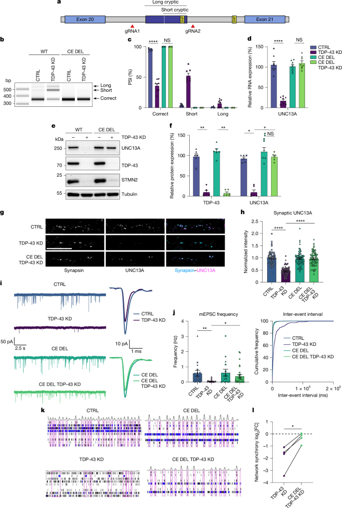
a, Schematic of genomic deletion of the UNC13A cryptic exon (CE). b–f, Analysis of WT and CE deletion (CE DEL) HaloTDP iNeurons on DIV28 following 14 days of HaloPROTAC treatment. b, RT–PCR showing that CE DEL restores UNC13A exon 20–21 splicing after TDP-43 knockdown. c, Quantification of b; n = 6 biological replicates from 3 experiments. d, RT–qPCR of correctly spliced UNC13A showing rescued RNA levels after TDP-43 knockdown; n = 6 biological replicates from 3 experiments. e, Western blot showing rescue of UNC13A protein levels by CE DEL. f, Quantification of e; n = 6 biological replicates from 3 experiments. g, Synapsin and UNC13A immunofluorescence in 4-week-old HaloTDP and CE DEL iNeurons cultured on rat astrocytes after 2 weeks of TDP-43 knockdown. h, UNC13A intensity at synapsin-positive terminals (control n = 71; TDP-43 knockdown n = 71; cryptic exon Del n = 66; CE DEL + TDP-43 knockdown n = 75 synapses; 3 experiments). i, Representative and mean mEPSC traces. j, mEPSC frequency and inter-event interval cumulative distributions (control n = 17; TDP-43 knockdown n = 22; CE DEL n = 15; CE DEL + TDP-43 knockdown n = 28 iNeurons; 3 experiments). k, Representative DIV39 MEA raster plots (60 s) for control, TDP-43 knockdown, CE DEL and CE DEL + TDP-43 knockdown; electrode bursts, blue; network bursts, pink. l, Network synchrony following TDP-43 knockdown, expressed as log2[fold change] in normalized electrode cross-correlation (n = 4 independent experiments); lines connect parallel differentiations recorded on the same MEA plate. Statistics: one-way analysis of variance (ANOVA) with Tukey’s test (c,d); repeated-measures one-way ANOVA on log-transformed, loading control-normalized values with Tukey’s test (f); Gaussian LMM with two-sided Satterthwaite-corrected t-tests (h,j); two-sided paired ratio t-test (l). P < 0.05; P < 0.01; P < 0.001; ****P < 0.0001. Scale bar, 10 µm. gRNA, guide RNA. Uncropped gels are shown in Supplementary Fig. 2.
UNC13A cryptic exon drives synapse dysfunction
TDP-43 knockdown yields synaptic transmission defects similar to those induced by direct depletion of UNC13A. However, TDP-43 has hundreds of validated RNA targets, regulating splicing, stability and transport of transcripts involved in virtually every aspect of neuronal function. Therefore, the reduction in synaptic transmission could reflect a multi-gene insult affecting pathways including synaptic vesicle trafficking, Ca2+ signalling and active zone organization. To determine the extent to which UNC13A cryptic exon inclusion contributes to impaired synaptic function following loss of TDP-43, we performed whole-cell patch clamping experiments in WT and cryptic exon deletion lines. Rescue of this single cryptic exon largely restored synaptic transmission following TDP-43 depletion with an 80% recovery in mEPSC frequency (Fig. 2i,j). mEPSC amplitude was still marginally (roughly 10%) reduced, supporting the conclusion that UNC13A cryptic exon deletion rescues specifically presynaptic function (Extended Data Fig. 7a). Passive membrane properties were similar between conditions (Extended Data Fig. 7b). As mEPSC frequency is influenced by synapse number, we measured excitatory synapse density along the dendrites of iNeurons. We found that TDP-43-knockdown neurons had fewer synapses than controls, a feature that persisted even when the UNC13A cryptic exon was deleted (Extended Data Fig. 7c,d), suggesting a strong recovery in synapse function in the remaining synapses.
Despite the small reduction in synapse number, MEA recordings showed that deletion of the UNC13A cryptic exon in TDP-43-depleted neurons restored synchronous network activity, indicating effective neurotransmitter release and propagation of signalling across the network (Fig. 2k,l and Extended Data Fig. 7e). This represented the most robustly rescued MEA metric (Extended Data Fig. 7f). Taken together, these findings show that correction of a single cryptic exon splicing event (intron 20 UNC13A cryptic exon) is sufficient to restore synaptic transmission and rescue the asynchronous patterns of network activity caused by TDP-43 loss. Active electrodes and cell coverage were comparable across conditions (Extended Data Fig. 7f,g). Likewise, analysis of neuronal survival confirmed that network-level changes were not attributable to changes in viability due to TDP-43 loss (Extended Data Fig. 7h) demonstrating that TDP-43 driven UNC13A loss drives a functional synaptic deficit in otherwise structurally intact, viable neurons.
UNC13A splice-switching ASOs
The rescue of UNC13A levels and restoration of synaptic signalling conferred by deletion of the UNC13A cryptic exon in the context of TDP-43 knockdown highlights the therapeutic potential of targeting this event in patients. We therefore sought to identify ASOs that can induce splice switching similar to those successful in targeting splicing events in SMN2 and STMN2 (refs. 23,24). We first designed locked nucleic acid-modified ASOs that targeted regions covering the cryptic exon acceptor and donor splice sites (Extended Data Fig. 8a). We screened the ASOs by transfection in SK-N-BE(2) cells and assessed their ability to restore UNC13A splicing at the exon 20–21 junction by RT–PCR (Extended Data Fig. 8b) and found that 21-nt ASOs targeting the donor splice site could restore UNC13A splicing. We confirmed splicing rescue and ability to restore UNC13A RNA and UNC13A protein levels in SH-SY5Y cells (Extended Data Fig. 8c–g). To improve therapeutic tolerance, we adapted the best performing ASOs to have full 2′-O-methoxyethyl (MOE) ribose-modified bases and tested their ability to rescue UNC13A splicing and restore protein levels in HaloTDP iNeurons (Fig. 3a–c and Extended Data Fig. 8h). We found these could restore splicing to more than 90% and protein levels to 30–50% of normal levels. Treatment with the M21-4 MOE ASO led to increased presynaptic UNC13A protein expression at synapsin-positive puncta following TDP-43 depletion compared with treatment with the control scrambled ASO (Fig. 3d). To identify ASOs that could increase protein rescue above 50% of normal levels, we carried out a secondary screen by performing an ASO walk along the cryptic donor site with 18-nt MOE-based ASOs (Extended Data Fig. 9a). We evaluated the ability of the 18-nt ASOs to rescue splicing in SH-SY5Y cells by RT–PCR (Extended Data Fig. 9b) and confirmed that the best performing 18-nt ASOs could rescue correct UNC13A mRNA splicing to more than 90%, and protein to more than 50% in SH-SY5Y cells and HaloTDP iNeurons (Fig. 3e,f and Extended Data Fig. 9c–h). In addition, we performed amplicon sequencing, which confirmed the ability of ASOs to rescue both reference and risk alleles in our HaloTDP iNeurons (Extended Data Fig. 9i).
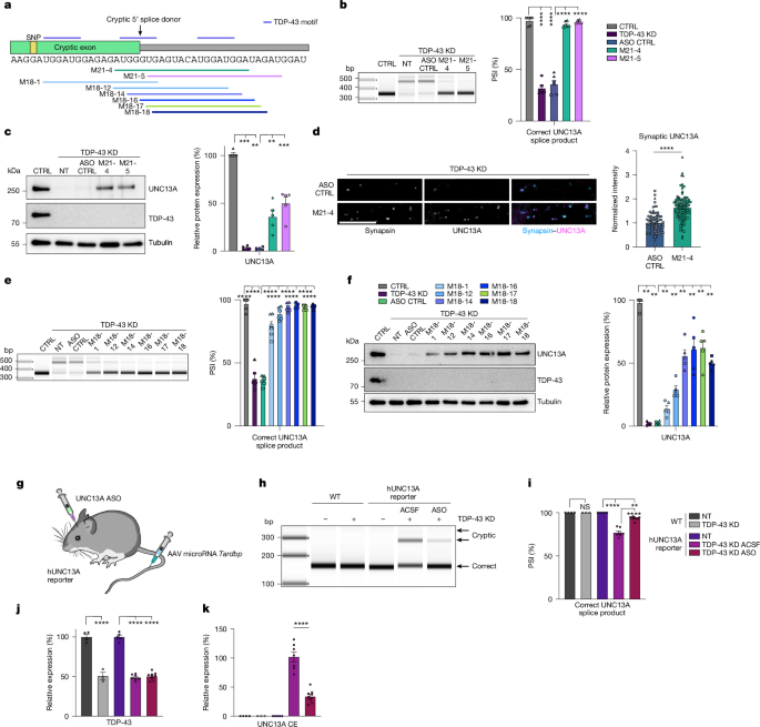
a, Schematic of ASOs targeting the UNC13A cryptic donor splice site. b, RT–PCR showing 21-nt ASOs prevent UNC13A cryptic splicing after TDP-43 knockdown, with quantification (percentage spliced in; n = 6 biological replicates from 3 experiments). c, Western blot showing rescue of UNC13A protein following 21-nt ASO treatment after TDP-43 knockdown, with quantification (n = 5 biological replicates from 3 experiments). d, UNC13A intensity at synapsin-positive terminals in TDP-43-knockdown iNeurons treated with MOE CTRL ASO (n = 88) or M21-4 ASO (n = 96). e, RT–PCR showing 18-nt ASOs prevent cryptic splicing after TDP-43 knockdown, with quantification (percentage spliced in; n = 6 biological replicates from 3 experiments). f, Western blot showing rescue of UNC13A protein following 18-nt ASO treatment after TDP-43 knockdown, with quantification (n = 5 biological replicates from 3 experiments). g, Schematic of the in vivo ASO splice correction experiment. h, RT–PCR of spinal cord from WT and hUNC13A reporter mice showing that M21-4 ASO reduces cryptic splicing after TDP-43 knockdown. i, Quantification of h (percentage spliced in). WT NT n = 4, WT TDP-43 KD n = 3, hUNC13A NT n = 5, hUNC13A ACSF n = 7, hUNC13A ASO n = 9. j,k, RT–qPCR of TDP-43 (j) and UNC13A cryptic exon (k) in WT and hUNC13A reporter mice. Statistics: one-way ANOVA with Tukey’s test (b,e,i–k); repeated-measures one-way ANOVA on log-transformed, loading control-normalized values with Tukey’s test (c,f); Gaussian LMM with two-sided Satterthwaite-corrected t-test (d). P < 0.01; P < 0.001; ***P < 0.0001. Scale bar, 10 µm. Uncropped gels are shown in Supplementary Fig. 3. ACSF, artificial cerebrospinal fluid; AAV, adeno-associated virus. Illustrations in g adapted from the NIH BioArt Source (https://bioart.niaid.nih.gov/bioart/505).
To validate the efficacy of UNC13A ASOs to rescue cryptic splicing in an in vivo model, we generated humanized UNC13A (hUNC13A) reporter mice, in which mouse intron 20 was replaced with human UNC13A intron 20 containing the cryptic exon. TDP-43 knockdown was performed by tail vein injection of an adeno-associated virus encoding a synthetic microRNA targeting the mouse Tardbp gene (Fig. 3g). On the same day, intracerebroventricular injections were performed in hUNC13A reporter mice with the M21-4 MOE ASO or artificial cerebrospinal fluid. Three weeks post-injection, spinal cords were dissected from the mice. RT–PCR analysis from spinal cord tissue following knockdown of TDP-43 showed no cryptic exon inclusion in WT mice and robust cryptic exon inclusion in hUNC13A reporter mice, which was partially rescued by administration of UNC13A splice-correcting ASO (Fig. 3h–k). Overall, these results show that ASOs can reduce inclusion of the UNC13A cryptic exon on TDP-43 knockdown both in vitro and in vivo, and can rescue UNC13A protein expression to above 50% in cultured neurons.
UNC13A ASOs rescue synapse function
Unc13a heterozygous knockout mice and individuals with heterozygous LoF UNC13A mutations are viable and do not present with abnormal neurophysiology18,25. Recovery of UNC13A expression to WT levels after TDP-43 LoF is probably not required to restore synapse function, in agreement with previous work in primary mouse cultures that showed that clear deficits in synapse function are only observed when expression of Unc13a falls below 30% of normal physiological levels20,25. Indeed, treatment with M18-17 ASO, which rescues UNC13A levels to more than 50% of control, was able to rescue synaptic transmission as measured by an increase in mEPSC frequency (Fig. 4a). We then tested our M21-4 ASO, which rescues UNC13A protein levels to roughly 30% of normal levels. Despite the lower level of protein rescue, treatment with M21-4 ASO also rescued synaptic transmission following TDP-43 depletion as measured by an increase in mEPSC frequency (Fig. 4b). mEPSC amplitude and passive membrane properties were unaffected by treatment with ASOs (Extended Data Fig. 10a–d), suggesting that ASO treatment specifically rescues presynaptic deficits caused by the cryptic exon in UNC13A.
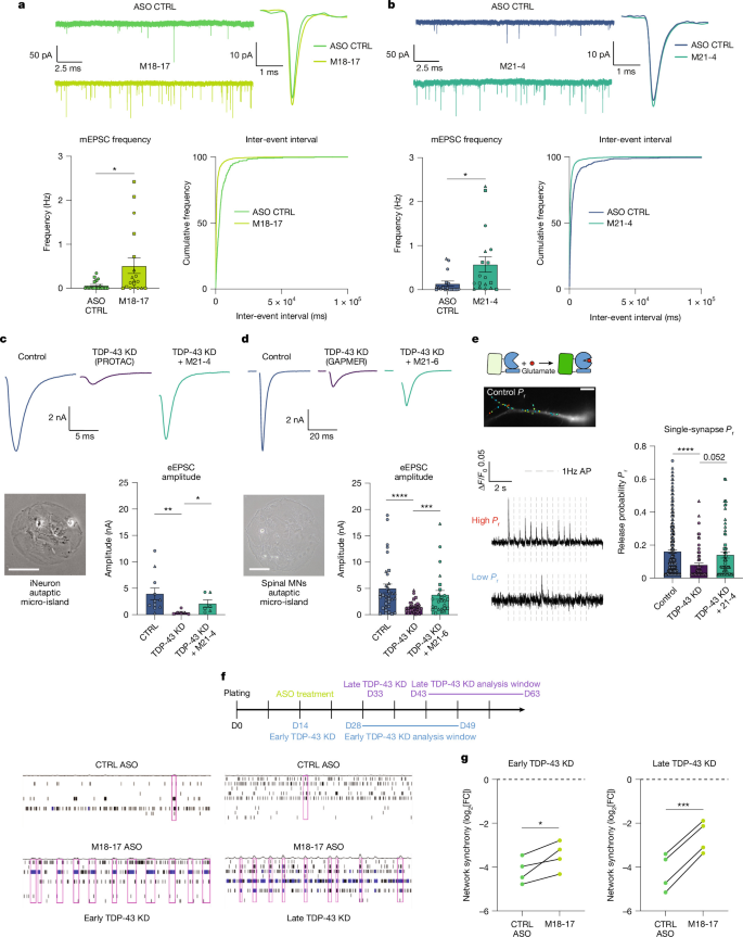
a, Representative and mean mEPSC traces from TDP-43-knockdown iNeurons treated with CTRL ASO (n = 19) or M18-17 ASO (n = 19), with quantification of mEPSC frequency and inter-event interval cumulative distributions. b, Representative and mean mEPSC traces from TDP-43-knockdown iNeurons treated with MOE CTRL ASO (n = 17) or M21-4 ASO (n = 18), with quantification of mEPSC frequency and inter-event interval cumulative distributions. c, Representative autaptic micro-island image and eEPSC traces from control, HaloPROTAC-mediated TDP-43 knockdown (DIV14) and TDP-43 knockdown + M21-4 ASO iNeurons, with eEPSC amplitude quantification (control n = 11; knockdown n = 6; knockdown + M21-4 n = 6; 3 independent differentiations). d, Representative autaptic micro-island image and eEPSC traces from control and gapmer-mediated TDP-43-knockdown (DIV45) spinal motor neurons (MNs) treated with M21-6, with eEPSC amplitude quantification (control n = 34; knockdown n = 30; knockdown + M21-6 n = 25 neurons; 3 independent differentiations). e, Example iNeuron dendrite expressing iGluSnFR3. Synapses were classified by release probability (Pr) after 45 field stimuli at 1 Hz. Quantification of single-synapse Pr in control (n = 285), TDP-43-knockdown (n = 71) and knockdown + M21-4 ASO (n = 66) synapses from 3 independent experiments. ΔF/F0, fractional change in fluorescence. f, Schematic of early and late TDP-43 knockdown in MEA recordings with representative raster plots; DIV49 for early, DIV45 for late (60 s); electrode bursts, blue; network bursts, pink. D, day. g, Network synchrony (log2[fold change versus control]) in HaloTDP iNeurons treated with CTRL or M18-17 ASO (n = 4 independent experiments); lines connect parallel differentiations recorded on the same MEA plate. Statistics: Gaussian LMM with two-sided Satterthwaite-corrected t-tests (a–e); two-sided paired ratio t-test (g). P < 0.05; P < 0.01; P < 0.001; ****P < 0.0001. Scale bars, 100 µm.
Whereas mEPSC recordings allowed us to detect deficits in spontaneous vesicle release, we also sought to determine whether TDP-43 depletion affected evoked synaptic transmission, the physiological mode that drives circuit function, and whether UNC13A ASOs could be beneficial. We conducted whole-cell voltage clamp recordings to examine evoked excitatory postsynaptic currents (eEPSCs) in autaptic micro-island cultures in which single neurons are grown on astrocytes and form synaptic connections onto themselves26,27. We performed experiments in two different ALS-affected neuronal subtypes: cortical-like excitatory iNeurons expressing the layer V marker CTIP2 and human iPS cell-derived spinal motor neurons expressing Islet-1 and the marker VAChT, performed in two laboratories with different methods for TDP-43 depletion (days in vitro (DIV) 14 PROTAC or DIV45 gapmer ASO knockdown). In both neuronal subtypes, TDP-43 depletion caused a substantial reduction in eEPSC amplitude, which was partially rescued by UNC13A splice-correcting ASOs (Fig. 4c,d and Extended Data Fig. 10e,f). Furthermore, the spontaneous vesicle release phenotype (decrease in mEPSC frequency) and ASO-mediated rescue were also confirmed in the separate spinal motor neuron model (Extended Data Fig. 10g–k). The amplitude of eEPSCs depends on several factors, including presynaptic probability of neurotransmitter release, postsynaptic strength (quantal amplitude) and synapse density. Our previous observations have already described UNC13A-independent decreases in both postsynaptic strength (Extended Data Fig. 7a) and synapse density (Extended Data Fig. 7c,d). Therefore, we decided to directly measure evoked presynaptic release probability at the level of single synapses in conventional iNeuron cultures (not autaptic), using live imaging of a genetically encoded reporter of glutamate release, iGluSNFR3 (ref. 28). TDP-43 depletion led to both a reduction in the number of active synapses as well as a reduction in the release probability of those synapses (Fig. 4e and Extended Data Fig. 11a). UNC13A splice-correcting ASOs did not rescue the number of active synapses (Extended Data Fig. 11a), consistent with the morphological analysis showing UNC13A splice correction also failed to rescue synapse density along dendrites following TDP-43 loss (Extended Data Fig. 7c,d and Extended Data Fig. 11b). However, UNC13A splice correction was associated with improved release probability at the remaining synapses, although this effect narrowly missed statistical significance (P = 0.052) (Fig. 4e).
Next, we tested the effectiveness of UNC13A splice-switching ASOs in rescuing network synchrony in neurons in which we induced TDP-43 depletion at DIV14. MEA recordings showed a small but statistically significant rescue in network synchrony following treatment with M18-17 ASO compared with a scrambled control (Fig. 4f,g and Extended Data Fig. 11c). To better mimic the disease, in which TDP-43 loss occurs in adult neurons, we induced TDP-43 depletion at DIV33, when neurons have already developed mature network activity (Extended Data Fig. 11d,e). As with earlier knockdowns, synchrony was the metric most affected by TDP-43 depletion (Extended Data Fig. 11f). In this case, ASO rescue experiments showed a greater recovery of network synchrony (Fig. 4f,g and Extended Data Fig. 11g). Across all MEA metrics, network synchrony showed the most consistent and pronounced rescue (Extended Data Fig. 11h).
Together, these data demonstrate that therapeutic targeting of the UNC13A cryptic exon using an antisense oligonucleotide can rescue synaptic function: from neurotransmitter release at individual synapses through to emergent network-level function.
Discussion
In this study, we show that TDP-43 LoF leads to a drastic loss of spontaneous and evoked synaptic transmission, leading to a disordered and asynchronous pattern of network activity in human neurons. Of the many hundreds of widespread cryptic mis-splicing events driven by TDP-43 LoF, we demonstrate that a single cryptic exon in the disease progression modifier gene UNC13A is a main driver of this synaptic breakdown. By designing splice-correcting ASOs targeting the UNC13A cryptic exon, we demonstrate how synaptic function can be restored, supporting UNC13A splice correction as a genetically anchored therapeutic strategy amenable to most patients with ALS and patients with FTD with TDP-43 pathology.
Out of the numerous cryptic splicing events induced by loss of nuclear TDP-43 that affect proteins that are predicted to affect neuronal health and function3,4,5,6,7,8,29, the cryptic exon in intron 20 of the UNC13A gene has attracted considerable interest. Risk SNPs accounting for the second strongest ALS and frontotemporal lobar degeneration with TDP-43 aggregation GWAS signal are located within this intron and enhance cryptic splicing, exacerbating transcript and protein loss6,7,14,15,16,30,31,32. Among patients with ALS, homozygous carriers of UNC13A risk SNPs (rs12608932 and rs12973192) show a 5–12-month reduction in median survival15,16,33,34,35,36. Therefore, genetic data strongly support targeting this event to modify disease progression.
The functional implications of UNC13A cryptic splicing in ALS and/or FTD remain unknown. UNC13A protein is crucial for synaptic vesicle priming; Unc13a-null mice die at birth from respiratory failure, and primary cortical cultures from these mice develop normally but have substantially reduced excitatory synaptic transmission18,19,20. Although ALS-linked mutations in TDP-43 have been shown to cause excitability and synaptic deficits in human neurons37,38,39, these studies did not establish a mechanistic role for cryptic exon inclusion or altered UNC13A protein levels. Therefore, we sought to determine whether direct depletion of TDP-43, modelling TDP-43 LoF in patients with sporadic ALS and associated alterations in RNA splicing also drive impaired synaptic function. Here we demonstrate TDP-43 depletion in iPS cell-derived neurons drives a reduction in both spontaneous and evoked synaptic vesicle release, leading to a failure of network-level signal propagation and asynchronous network activity. These results mirror the LoF we observed following direct UNC13A knockdown, as well as the synaptic defects described in previously published reports of Unc13a depletion in primary rodent neurons18,19,20.
TDP-43 LoF causes widespread mis-splicing in hundreds of transcripts, many of which are involved in synaptic transmission. Therefore, to specifically assess the contribution of the UNC13A cryptic exon to the decreased synaptic activity observed in TDP-43 depleted neurons, we selectively deleted the UNC13A intron 20 cryptic exon in human neurons. We found that rescuing this single mis-splicing event could restore spontaneous and evoked synaptic vesicle release in two independent models (cortical-like iNeurons and spinal motor neurons), and re-establish network synchrony, demonstrating that loss of UNC13A protein is a main driver of synaptic breakdown caused by TDP-43 LoF. Other synaptic genes are also likely to contribute to the broader range of observed phenotypes. For example, we observed changes in mEPSC amplitude on TDP-43 loss in iNeurons that are not recapitulated by direct UNC13A loss, nor rescued by deletion of the UNC13A cryptic exon. Similarly, we observed a reduction in synapse density following TDP-43 knockdown that does not appear to be driven by altered UNC13A protein levels. Further studies, focusing on mis-spliced transcripts whose products either localize to the postsynapse or are involved in synapse turnover40, will explain the functional mechanisms underpinning these changes. Despite this strong deficit in synapse density that is independent of UNC13A, we found that rescuing UNC13A led to a robust recovery in both spontaneous and evoked synaptic transmission as measured across two independent model systems and cell types relevant to ALS and/or FTD (cortical-like iNeurons and spinal motor neurons). Live imaging of individual synapses revealed that, although the number of active synapses was not restored, there was a trend towards higher release probability of these remaining synapses after splice correction, highlighting the importance of vesicle release kinetics at individual synapses in contributing to emergent network-level function.
Data assessing UNC13A protein levels in patient neurons are not available, but transcriptomic data on fluorescence-activated nuclei-sorted postmortem cortex from patients with ALS and/or FTD with TDP-43 pathology show that the UNC13A cryptic exon can be included in more than 90% of UNC13A transcripts within the cell population lacking nuclear TDP-43, which is thus predicted to greatly inhibit production of functional UNC13A protein6,41. These findings support the validity of using iPS cell-derived neuronal models in which TDP-43 loss induces a near-total loss of UNC13A, although models that recapitulate both nuclear loss and cytoplasmic aggregation will be beneficial in future work. Previous electrophysiology studies have demonstrated that the level or activity of UNC13A needs to be reduced below roughly 30% before strong synaptic deficits appear20,25. This agrees with studies using Unc13a-heterozygous mice, which showed no defects in synaptic activity. Our results show that restoring roughly 30% or more of UNC13A protein levels can rescue synaptic transmission. Altogether, this suggests that splice-switching therapeutic rescue of UNC13A to more than 30% in patients with ALS could be beneficial for the recovery of synaptic function.
Although loss of UNC13A effectively leads to synaptic silencing, transcranial magnetic stimulation studies have detected cortical hyperexcitability at an early stage in patients with ALS that worsens with disease progression42 and is associated with poorer survival outcomes43. Although other factors, such as KCNQ2 mis-splicing, may contribute to this hyperexcitability, it is possible that UNC13A loss also plays a role in this. Rare patients with inherited biallelic UNC13A mutations can present with seizures at a young age25,44,45, and one individual with a biallelic truncating mutation in UNC13A developed myasthenia and had cortical hyperexcitability on electroencephalography analysis44. Cortical hyperexcitability has also been shown in individuals with variants in other presynaptic proteins that reduce synaptic vesicle release through LoF or dominant-negative mechanisms, such as STX1B (syntaxin-1B)46, STXBP1 (Munc18-1)47 and SNAP25B (ref. 48). The impaired excitatory synaptic output driven by UNC13A mis-splicing could drive hyperexcitability through more complex circuit alterations, such as the reduction in microcircuit feedforward inhibition and an overall skew of the excitatory-to-inhibitory ratio49. Our experimental system consisted exclusively of excitatory neurons, in which impaired synaptic transmission manifested as a loss of network synchrony, rather than hyperactivity. Future studies should incorporate inhibitory interneurons to more precisely assess how TDP-43 loss and UNC13A dysfunction affect network dynamics. The humanized mouse model described here could provide a valuable platform for assessing network changes in an in vivo setting.
Our experiments were carried out in populations of neurons that had a homogeneous loss of TDP-43 and in which its presynaptic and postsynaptic effects are inevitably compounded. As such, our experimental setup represents a more extreme condition than the mosaic pattern of TDP-43 loss seen in patients with ALS, in which affected neurons can still project onto unaffected neurons. It is conceivable that UNC13A-mediated presynaptic release rescue could be sufficient to restore normal synaptic signalling in connections in which the postsynaptic neuron remains unaffected, and future studies in more complex models will help resolve how circuits are affected by mosaic UNC13A mis-splicing.
The cellular events that lead to TDP-43 aggregation in ALS are unknown and it is therefore challenging to correct this directly, but aggregation is associated with a loss of TDP-43 nuclear function that produces numerous cryptic splicing events, which may represent more tractable therapeutic targets than targeting mislocalization directly. Splice-modifying ASOs have been successfully developed to treat spinal muscular atrophy by correcting SMN2 splicing23,50. This same strategy can be used to correct cryptic splicing in ALS and has the advantage of being able to restore physiological transcript levels, without the risk of overexpression. Yet there is still a need to identify splicing events that have the strongest impact on neuronal function and hold the most promise of improving disease symptoms. Our findings, along with results from GWAS suggesting that modulation of UNC13A cryptic exon inclusion affects disease progression15, strongly support UNC13A as a key candidate for splice-modifying therapy development in ALS. TDP-43 LoF drives sufficient UNC13A reduction to impair synaptic transmission and network synchrony.
We show prevention of UNC13A mis-splicing by means of CRISPR–Cas9 gene editing or application of ASOs can rescue UNC13A protein expression and restore defects at many layers of complexity: from single-synapse release probability to spontaneous and evoked synaptic transmission and emergent network synchrony. This points to a stage of TDP-43-linked neurodegeneration in which neurons lose the ability to contribute to meaningful synaptic communication before overt neuronal death. This functional death provides a unique therapeutic opportunity: a pool of structurally intact neurons unable to contribute to normal network function that are recoverable through splice-correcting ASOs targeting a single TDP-43 dependent cryptic exon in UNC13A. Notably, therapies targeting UNC13A splicing would be amenable to most patients with ALS and/or FTD and TDP-43 pathology that is supported by clear genetic evidence and a clearly defined molecular and functional mechanism.
Methods
Cell culture (University College London)
Key reagents can be found in Supplementary Table 1. SH-SY5Y cells (American Type Culture Collection (ATCC)) and SK-N-BE(2) (ATCC) with stable doxycycline-inducible short-hairpin RNA (shRNA) to TARDBP were described in ref. 6 and were maintained in DMEM–F12 with GlutaMAX (Thermo Fisher Scientific) with 10% fetal bovine serum (FBS) (Thermo Fisher Scientific), 1% penicillin–streptomycin (Thermo Fisher Scientific) and split with TrypLE Express (Thermo Fisher Scientific) when 80% confluent. The WTC11 iPS cell line derived from a healthy human participant (GM25256) with stable integration of doxycycline-inducible expression of NGN2 at the AAVS1 locus and stable integration of dCas9-BFP-KRAB at the CLYBL locus was a gift from M. Ward51. This iPS cell line is heterozygous for UNC13A risk SNPs (rs12608932 and rs12973192). Cells were maintained in E8 Flex Medium (Thermo Fisher Scientific) in Geltrex-coated (Thermo Fisher Scientific) plates and passaged with Versene solution (Thermo Fisher Scientific) or Accutase (Thermo Fisher Scientific) when 80% confluent. All cell lines were authenticated by short tandem repeat profiling and were routinely tested for mycoplasma contamination, with negative results throughout. For induction of iPS cells to iNeurons, iPS cells were passaged with Accutase (Thermo Fisher Scientific) and plated into Geltrex-coated plates with induction media: DMEM–F12 with GlutaMAX (Thermo Fisher Scientific), 1× N2-Max Supplement (R&D Systems), 1× non-essential amino acids (Thermo Fisher Scientific), 2 μg ml−1 doxycycline hyclate (Sigma-Aldrich), 2 μM XAV939 (Cayman Chemical), 10 μM SB431542 (Cayman Chemical) and 100 nM LDN-193189 (Cayman Chemical)52,53. Medium was changed daily for 3 days. On the third day, cells were replated to poly-l-ornithine-coated (10 μg ml−1; Sigma-Aldrich) plates for RNA and protein experiments or on 50 μg ml−1 poly-d-lysine (PDL) (Gibco; Thermo Fisher Scientific) and 10 μg ml−1 laminin-coated (Gibco; Thermo Fisher Scientific) 18-mm coverslips (Deckglaser) with primary rat astrocytes for electrophysiology, live imaging and immunofluorescence experiments. Differentiated neurons were maintained in neuronal medium (synaptic medium) with extra supplements: BrainPhys (StemCell Technologies), with 1× N2-Max Supplement (R&D Systems), 1× N21-Max Supplement (R&D Systems), 10 ng ml−1 NT3 (StemCell Technologies), 20 ng ml−1 glial cell line-derived neurotrophic factor (GDNF) and brain-derived neurotrophic factor (BDNF) (Peprotech), 2.5% FBS (Thermo Fisher Scientific), 1× Non-Essential Amino Acids (Thermo Fisher Scientific), 0.2 mg ml−1 cAMP (Sigma-Aldrich), 1× GlutaMAX (Thermo Fisher Scientific), 1% penicillin–streptomycin (Thermo Fisher Scientific) and 200 nM ascorbic acid (Sigma-Aldrich)54. This medium was used for all experiments, with the exception of MEA recordings that were carried out without FBS. Half-medium changes were performed twice per week for all cultures, or after recording on MEA cultures.
For rat astrocyte preparation, primary cortices were dissected from E18 Wistar rat embryos (Charles River Laboratories) and treated with 0.5 mg ml−1 trypsin (Worthington) for 15 min at 37 °C. Cortices were washed with Hanks’ balanced saline solution (Thermo Fisher Scientific) and triturated with fire-polished glass Pasteur pipettes of decreasing diameter in DMEM (Thermo Fisher Scientific) + 10% FBS (Gibco; Thermo Fisher Scientific). Cells were collected by centrifugation at 900 r.p.m. for 10 min, resuspended and passed through a 70-µm cell strainer. Roughly 15–18 million cells were plated in a T-75 flask and cultured in DMEM + 10%FBS + 1% penicillin–streptomycin (Thermo Fisher Scientific). After 2 days a complete medium change was conducted to remove antibiotics and medium was replaced every 3 days thereafter. Once reaching confluence, the cultures were enriched for astrocytes by shaking at 180 r.p.m. on an orbital shaker for 30 min, after which the medium was removed. Fresh medium was added and shaking increased to 240 rpm for 6 h, after which the medium was replaced. These steps remove microglia and oligodendrocyte precursor cells. Astrocyte-enriched cultures were passaged using 0.25% trypsin–EDTA (Thermo Fisher Scientific) and cultured in fresh T-75 flasks for 12–14 days, with a medium change every 2–3 days, until confluent. Astrocytes were then passaged and either stored in liquid nitrogen or plated on 50 μg ml−1 PDL (Gibco; Thermo Fisher Scientific) and 10 μg ml−1 laminin-coated (Gibco; Thermo Fisher Scientific) 18 mm coverslips (Deckglaser) for culturing with iNeurons.
Cell culture (Leibniz-FMP-Berlin)
The GM25256 human iPS cell line, which is heterozygous for UNC13A risk SNPs (rs12608932 and rs12973192) (Coriell Institute for Medical Research) was maintained in mTeSR1 stem cell culture media on Matrigel-coated plates (StemCell Technologies, 85850; Corning, 354277). Human iPS cells were fed daily and split every 4–5 days. Differentiation of iPS cells into motor neurons (iMNs) was carried out using three-dimensional developmental pathways for 14 days (refs. 7,55). On day 14, neuronal spheroids were dissociated, counted and frozen using 70% KnockOut Serum Replacement (Gibco; Thermo Fisher Scientific, 10828010), 20% 1 M Trehalose (Thermo Fisher Scientific, 182550250) and 10% DMSO (Sigma-Aldrich, D2650). Neurons were thawed, and plated on PDL–laminin-coated coverslips or directly on PDL-coated coverslips on which mouse astrocytes were plated25, and maintained in a medium containing Neurobasal medium (Gibco; Thermo Fisher Scientific, 12348017) supplemented with 1× N-2 supplement (Gibco; Thermo Fisher Scientific, 17502001), 1× B-27 supplement (Gibco; Thermo Fisher Scientific, A1486701), 1× penicillin–streptomycin (Gibco; Thermo Fisher Scientific, 15140-122), 20 µM l-ascorbic acid (Sigma-Aldrich, A4403-100MG), 1% KnockOut Serum Replacement (Gibco; Thermo Fisher Scientific, 10828010), 5 µM ROCK inhibitor Y-27632 (StemCell Technologies, 72304), 10 ng ml−1 GDNF (R&D Systems, 212-GD-050), 10 ng ml−1 CNTF (R&D Systems, 257-NT-050) and 20 ng ml−1 BDNF (R&D Systems, 11166-BD-050) for the first 2 days. After 2 days, half of the medium was exchanged with neuron maintenance medium without the ROCK inhibitor and supplemented with 5 µM DAPT (Sigma-Aldrich, D5942) and 10 ng ml−1 BDNF. Five days later, iMNs were fed with neuron maintenance medium without ROCK inhibitor and DAPT. iMNs were supplemented with fresh medium every 7 days.
Spinal iMNs release the neurotransmitter acetylcholine, but do not express postsynaptic nicotinic receptors. As such, iMN cultures express synaptic markers, but the cells are not synaptically connected. To record synaptic transmission properties in these neurons, we expressed the vesicular glutamate transporter (VGlut1) tagged by green fluorescent protein (GFP) in iMNs 1 week before electrophysiological recordings, on the basis of previous observations that its expression can confer the release of glutamate in inhibitory neurons56. Complementary DNA (cDNA) encoding rat VGlut1 tagged with enhanced GFP (EGFP) at the carboxy terminus was used to prepare lentiviral particles (Viral Core Facility of the Charité–Universitätsmedizin Berlin). The FUGW vector57, in which the ubiquitin promoter was exchanged with the human synapsin 1 promoter, was used according to a previously published57, modified protocol58, using helper plasmids59 (Addgene, 8454 and 8455).
Lentivirus production for CRISPRi knockdown
sgRNA pU6-sgRNA EF1Alpha-puro-T2A-BFP vector (Addgene 60955) was used to express the following sgRNAs:
sgCTRL, GTCCACCCTTATCTAGGCTA
sgTARDBP, GGGAAGTCAGCCGTGAGACC
sgUNC13A, GGAACCAAGATGGCCGGTGG
sgRNA transfer plasmid with packaging (pCMVΔ8.91) and VSVG envelope (pMD.G) plasmids were transfected into human embryonic kidney 293T (HEK293T) cells (Horizon Discovery) with TransIT-293 (Mirus). Medium was collected on days 2 and 3 after transfection, combined and concentrated 1:10 with Lenti-X concentrator (Takara Bio).
HaloTag editing of TARDBP
iPS cells were electroporated with 10 μg of HaloTag-TARDBP homology-directed repair template (gift from M. Ward, National Institutes of Health (NIH)), 500 pmol of Cas12 CRISPR-RNA (crRNA) (GGAAAAGTAAAAGATGTCTGAAT, Integrated DNA Technologies) and 20 μg of recombinant Cas12 (Integrated DNA Technologies) using the P3 Primary Cell 4D-Nucleofector kit (Lonza) and 4D-Nucleofector X unit (Lonza) using the CA-137 program. After electroporation iPS cells were plated in E8 Flex Media supplemented with 1× RevitaCell (Thermo Fisher Scientific) and 1× Alt-R HDR Enhancer V2 (Integrated DNA Technologies) for 24 h and then expanded in E8 Flex Media. Cells were labelled with HaloTag-TMR ligand (Promega) and positive clones were selected for genotyping by PCR.
UNC13A exon 20–21 cryptic exon deletion
sgRNAs were designed to target 45 bp upstream of the acceptor splice site of the long cryptic exon (GGAAAAGTAAAAGATGTCTGAAT) and 10 bp downstream of the donor splice site (GAGATGGGTGAGTACATGGA). Sense and antisense bridging oligonucleotides 5′-AAAGTCCAGATTCAGGG TCACCTCCTCTGGGAAGCCCACC TTGGCCTCCBGGATGGATAGATGGATGAGTTGGTGGGTAGATTCGTGGCTAGATGGATGA-3′ and 5′-TCATCCATCTAGCCACGAATCTACCCACCAACTCATCCATCTATCCATCDTGGAGGCCAAGGTGGGCTTCCCAGAGGAGGTGACCCTGAATCTGGACTTT-3′, which include degenerate base B (C, G or T) and D (A, G or T) were designed as homology-directed repair templates. The WTC11 iPS cell line (GM25256) and SH-SY5Y were electroporated with 250 pmol of each Cas9 sgRNA (Integrated DNA Technologies) along with 20 μg of recombinant Cas9 (Integrated DNA Technologies) and 1 pmol of each bridging oligonucleotide using the P3 Primary Cell 4D-Nucleofector kit (Lonza) using the CA-137 program60. Following electroporation cells were maintained in media with 1× Alt-R HDR Enhancer V2 (Integrated DNA Technologies) for 24 h.
ASO synthesis
Locked nucleic acid-modified ASOs were purchased from Integrated DNA Technologies. The 21-nt MOE-modified UNC13A splice-correcting ASOs or TDP-43 gapmer ASOs were purchased from Dharmacon, Wuxi or Biospring, and a gift from Trace Neuroscience. A panel of 18-nt MOE-modified ASOs was designed to tile across the region of interest before being subjected to further selection on the basis of potential sequence-driven off-target effects and secondary structure. The resulting panel of 24 ASOs was generated by solid-phase synthesis using MerMade12 automated synthesizer using prepacked controlled pore glass columns (1-µmol scale, 1,000 Å). Phosphoramidites (TheraPure by Thermo Fisher Scientific) were prepared at 0.08 M using acetonitrile. Cleavage and deprotection of oligonucleotides were performed using concentrated ammonia for 6 h at 55 °C. After deprotection, samples were dried and resuspended in water. Purification of the oligonucleotides was performed by reversed-phase high-performance liquid chromatography using 0.1 M triethylammonium acetate pH 7 and acetonitrile in an XBridge BEH C18 Prep column (Waters). Oligonucleotides were desalted using ethanol precipitation and quantification performed measuring the absorbance at 260 nm and the corresponding molecular extinction coefficient for each ASO. Quality control of the synthesized oligonucleotides was performed by liquid chromatography with mass spectrometry. ASO sequences are in Supplementary Table 2.
ASO treatment (University College London)
SH-SY5Y cells with stable integration of a shRNA cassette for TARDBP were treated with 25 ng ml−1 doxycycline (Sigma-Aldrich) for 5 days and then transfected while replating into 12-well plates using Lipofectamine RNAiMax (6 μl per well, Thermo Fisher Scientific) and ASOs at 50 nM final concentration. Following transfection, doxycycline concentration was increased to 1 μg ml−1 and cells were collected 5 days after transfection. To test 18-nt MOE-based ASOs, shTARDBP SH-SY5Y cells were treated with 25 ng ml−1 doxycycline for 5 days, then 100 ng ml−1 doxycycline for 24 h, plated into 96-well plates and transfected using Lipofectamine RNAiMax (0.72 μl per well) and ASOs at 10 nM or 50 nM final concentration. Cells were collected 48 h after transfection.
SK-N-BE(2) cells with stable integration of a shRNA cassette for TARDBP were treated with 25 ng ml−1 doxycycline for 3 days and then transfected while replating into 12-well plates using Lipofectamine RNAiMax (6 μl per well) and ASOs (4 μl of 100 μM stock). Following transfection doxycycline concentration was increased to 1 μg ml−1 and cells were collected 3 days after transfection.
HaloTDP iNeurons were cultured for 2 weeks after replating in neuronal media and then treated with 30 nM HaloPROTAC-E21 (University of Dundee) or HaloPROTAC3 (Promega). ASOs were added directly to neuronal media to enable free uptake. ASO treatments (1 μM) for protein and RNA collection were started at the same time as PROTAC treatment on DIV14 and were included in each media change for 2 weeks until cells were collected. ASO details for all functional experiments are outlined in Supplementary Table 3.
ASO treatment (Leibniz-FMP-Berlin)
To make 100 µg µl−1 storage stock solution, lyophilized ASO was resuspended in the appropriate amount of sterile PBS, pH 7.4. Once fully dissolved, the ASO was filter sterilized using a spin column (0.22 µm). The 100 µg µl−1 storage stock solution of ASO was aliquoted and stored at −20 °C for future use. To make roughly 500 µM working stock solutions for dosing neurons, the 100 µg µl−1 storage stock solution was diluted appropriately with sterile PBS, pH 7.4 and filter sterilized using a spin column (0.22 µm). To determine the concentration of the ASO working stock solution, the absorbance at 260 nm was measured on a NanoDrop One (Thermo Fisher Scientific) and concentrations were calculated using the molecular extinction coefficient for that ASO. The working stock solution of ASO was aliquoted and stored at −20 °C for future use. Dilutions of working stocks to the appropriate dose levels were made with the appropriate amount of sterile PBS, pH 7.4. On day 45 after neuronal plating, TRTDP43 ((moeA)(moeA)(moeG)(moeG)(moe5C)(dT)(dT)(d5C)(dA)(dT)(dA)(dT)(dT)(dG)(dT)(moeA)(moe5C)(moeT)(moeT)*(moeT); where moe denotes MOE base, d denotes DNA base, 5C denotes 5-methylcytosine) (2–4 μM)) was added to the culture wells to knock down TDP-43. In parallel, UNC13A splice-correcting M21-6 (2–4 μM) was added to some of the samples. Application was carried out once a week for 3 weeks. Electrophysiological recordings were carried out precisely 1 week following the last application.
RT–qPCR
RNA was extracted from SH-SY5Y cells, SK-N-BE(2) cells and iNeurons with a RNeasy kit (Qiagen) using the manufacturer’s protocol including homogenization of samples with QIAshredder spin columns (Qiagen). RNA concentrations were measured by NanoDrop (Thermo Fisher Scientific) and 500 ng of RNA was used for reverse transcription. First-strand cDNA synthesis was performed with RevertAid (Thermo Fisher Scientific, K1622) using random hexamer primers and following the manufacturer’s protocol including all optional steps. Gene expression analysis was performed by RT–qPCR using TaqMan Multiplex Universal Master Mix (Thermo Fisher Scientific, 4461882) with custom IDT RT–qPCR assays (Supplementary Table 4) and TaqMan assays (GAPDH-Jun assay; Thermo Fisher Scientific, 4485713) on a QuantStudio 5 Real-Time PCR system (Applied Biosystems) and quantified using the ΔΔCt method61. For RT–qPCR based on intercalating dyes PowerUp SYBR Green master mix (Thermo Fisher Scientific) was used with gene-specific primers (Supplementary Table 4).
RT–PCR
RNA extraction and cDNA synthesis were performed as in RT–qPCR methods. The UNC13A cryptic exon was amplified with a forward primer in exon 19 5′-CAGACGATCATTGAGGTGCG-3′ and reverse primer in exon 22 5′-ATACTTGGAGGAGAGGCAGG-3′ using Q5 Hot Start High Fidelity 2× Master Mix (New England Biolabs). PCR products were resolved on a TapeStation 4200 (Agilent) and bands were quantified with TapeStation Systems Software v.3.2 (Agilent). For the primary screen of ASOs in SK-N-BE(2) cells, PCR products were analysed with a QIAxcel capillary electrophoresis machine (Qiagen). Raw data were exported and analysed with the QIAxcelR package (v.0.1) (https://github.com/Delayed-Gitification/QIAxcelR).
Amplicon sequencing
Genomic DNA from SH-SY5Y cells and iPS cells were extracted with the Monarch Genomic DNA Purification Kit (New England Biolabs). PCR was performed with Q5 Hot Start High Fidelity 2× Master Mix (New England Biolabs) and purified with Monarch PCR cleanup kit (New England Biolabs). Purified amplicons were sequenced using the Premium PCR long-read sequencing service (Plasmidsaurus). Raw FASTQ reads were mapped to UNC13A and TARDBP genes using Minimap2 (ref. 62) with the -x splice flag enabled (v.2.26-r1175) and converted to BAM files with SAMtools63 (v.1.21). Results were visualized with the Integrative Genomics Viewer (IGV) browser64. For quantification of UNC13A allele-specific rescue, RT–PCR was performed as described above and amplicons were sequenced by Plasmidsaurus. Raw FASTQ reads were mapped to UNC13A using Minimap2, converted to BAM files with SAMtools and SNPs were quantified with custom Python scripts.
Low-pass whole-genome sequencing
Genomic DNA was extracted from iPS cells using the Monarch Genomic DNA Purification Kit (New England Biolabs). Low-pass whole-genome sequencing (roughly 10× mean coverage) was performed by Azenta Life Sciences. Adapter trimming and quality control of FASTQ files were carried out with fastp65 (v.0.23.4). Reads were aligned to the human reference genome GRCh38 (GCA_000001405.15) using bwa-mem2 (ref. 66) (v.2.2.1), and the resulting alignments were converted to sorted BAM format with SAMtools (v.1.21). Copy-number variant analysis was performed with CNVkit67 (v.0.9.12) using the batch command with the whole-genome sequencing pipeline (--method wgs). Sequencing-accessible regions were defined by masking gaps and repeat tracks from the UCSC genome browser (--access access-hg38.bed). Bins of 20-kilobase average size were used (--target-avg-size 20,000), GC bias and low-coverage bins were corrected (--drop-low-coverage), gene annotations were added from refFlat.txt (--annotate) and a flat male reference was constructed (--male-reference --normal --output-reference WTC11-WT_flat.cnn). Segmented copy number profiles were generated with the default circular binary segmentation algorithm.
Off-target PCR and sequencing
For Cas9 genome edits (UNC13A cryptic exon deletion) off-target sites were identified using the CRISPR–Cas9 gRNA checker tool (Integrated DNA Technologies). For Cas12 genomic edits (HaloTag-TDP-43), off-target sites were predicted by Cas-OFFinder68 using the following settings: LB Cas12, PAM motif TTTV, DNA Bulge = 1 and mismatch = 3. Predicted off-targets sites within annotated genes were examined. Genomic DNA from iPS cells was extracted with Monarch Genomic DNA Purification Kit (New England Biolabs). PCR was performed with 50 ng of DNA using Q5 Hot Start High Fidelity 2× Master Mix (New England Biolabs) with the primers described in Supplementary Table 5.
PCR products were resolved on a TapeStation 4200 (Agilent) and bands were analysed with TapeStation Systems Software v.3.2 (Agilent). PCR products were sequenced using the Premium PCR long-read sequencing service (Plasmidsaurus) to generate full-length reads of individual amplicons. Raw FASTQ files were aligned with Minimap2, outputs were converted to BAM files with SAMtools (v.1.21) and then visualized with IGV.
Western blot analysis (University College London)
Cells were collected with RIPA buffer (Thermo Fisher Scientific) with cOmplete mini protease inhibitors (Sigma-Aldrich) and protein concentration was quantified using the Pierce bicinchoninic acid (BCA) Protein Assay Kit (Thermo Fisher Scientific). Protein lysates were resolved on 4–12% precast Bis-Tris gels (Thermo Fisher Scientific) or poured 7% Bis-Tris gels and transferred to 0.2-μm polyvinylidene difluoride membranes (Merck Millipore). Membranes were blocked with 5% non-fat milk in tris-buffered saline with Tween (TBST) and then probed overnight with primary antibodies (Supplementary Table 6) in 5% non-fat milk in TBST at 4 °C. Blots were washed 3 times with TBST and subsequently probed with horseradish peroxidase (HRP)-conjugated secondary antibodies at room temperature for 1 h (Goat anti-Mouse HRP Bio-Rad 1706516; 1:10,000 or Goat anti-Rabbit HRP Bio-Rad 1706515; 1:10,000) and developed with Chemiluminescent substrate (Merck Millipore, WBKLS0500) on a ChemiDoc Imaging System (Bio-Rad). Band intensity was measured with Fiji (ImageJ)69 (NIH version 2.0.0-rc-69). Band intensities were normalized to loading control (Tubulin) within each lane. Normalized values were log10-transformed before analysis. The effect of treatment was assessed by repeated-measures one-way analysis of variance (ANOVA), with biological replicate (blot) as the matching factor and the Geisser–Greenhouse correction applied (sphericity not assumed), followed by Tukey’s multiple comparisons test for all pairwise comparisons between groups.
Western blot analysis (Leibniz-FMP-Berlin)
Motor neurons were lysed in RIPA buffer (25 mM Tris, 150 mM NaCl, 0.5% (w/v) SDOC, 1% IGEPAL (NP-40), 0.1% (w/v) SDS) with 0.1% of each protease inhibitor (aprotinin, leupeptin, phenylmethylsulfonyl fluoride), sonicated and centrifuged in a tabletop centrifuge at 4 °C for 45 min at 15,000 r.p.m. Supernatants were transferred into a fresh tube, and protein concentration was determined using Pierce BCA Protein Assay Kit (Thermo Fisher Scientific, 23225). Samples were diluted in Laemmli buffer (3% (w/v) SDS, 41.6 mM Tris, 1 mM EDTA, 10% (v/v) glycerol, 0.06% (w/v) bromophenol blue, 0.77%, (w/v) dithiothreitol in double-distilled H2O (ddH2O)) and incubated for 10 min at 95 °C. Then 10 µg of the lysed samples was loaded onto a commercial 4–12% Bis-Tris gels (NuPAGE 4–12% Bis-Tris Gel (1.0 mm × 10 wells), Invitrogen, NP0321BOX) and separated in cold MOPS buffer (20 mM MOPS free acid, 5 mM sodium acetate, 1 mM Na2EDTA) at a constant voltage of 120 V. Transfer of the proteins was conducted in transfer buffer (192 mM glycine, 25 mM Tris, 10% methanol) on a nitrocellulose membrane for 14 h at 65 mA. Membranes were then stained for total protein as a loading control with Pierce Reversible Protein Stain Kit for Nitrocellulose Membranes (according to the manufacturer’s manual; Thermo Fisher Scientific, 24580). The membranes were blocked with 5% milk in TBST buffer (10 mM Tris/HCl, 100 mM NaCl, 0.1% Tween 20, pH 7.5) for 1 h at room temperature. After this step, the membranes were cut and incubated with primary antibodies: rabbit anti-Munc13-1 (Synaptic Systems, 126 103, 1:250), rabbit-TDP-43 (Proteintech, 10782-2-AP, 1:1,000), mouse anti-Vinculin (Sigma-Aldrich, V9131-100UL 1:1,000) and rabbit anti-Stathmin-2 (Bio-Techne, NBP1-49461, 1:1,000). The corresponding secondary antibodies used were: anti-mouse HRP (Jackson ImmunoResearch, 115-035-146, 1:3,000) and anti-rabbit HRP (Jackson ImmunoResearch, 111-035-144, 1:3,000). The chemiluminescence signal was detected with Pierce ECL Western Blotting Substrate (Thermo Fisher Scientific, 32106) on an ECL ChemoStar imager (Intas). Protein signals were analysed in Fiji (ImageJ). Protein levels were first normalized to the total protein staining in the corresponding lane, and then to the control condition. Finally, statistical analysis was conducted in GraphPad Prism (GraphPad Software) using one-way ANOVA with multiple comparisons. Error bars represent mean ± s.e.m.
MEA recordings
Maestro 96-well cytoview MEA plates (Axion Biosystems M768-tMEA-96B) were drop-wise coated in 0.05 mg ml−1 PDL (Gibco; Thermo Fisher Scientific, A38904-01) at room temperature for 2 h, washed 3× in 50 μl of ddH2O and left to dry overnight. Wells were then drop-wise coated with 10 μg ml−1 laminin (Thermo Fisher Scientific) for 1–2 h at 37 °C. 50,000 neurons and 12,500 primary rat astrocytes were drop-wise plated in synaptic media (described above) containing ROCK inhibitor (Y-27632; Tocris, 1254) at 1 μM. Cells were left to attach for 1.5 h before media containing CultureOne Supplement (Gibco; Thermo Fisher Scientific) was added for 3 days. Half-medium changes (without ROCK inhibitor or CultureOne) were thereafter performed every 2–3 days and HaloPROTAC-E 30–300 nM was added to cultures with each media change starting on DIV14 for early TDP-43 KD and at DIV33 for late TDP-43 KD. ASO treatment was performed every 2 weeks starting at DIV14, at a final concentration of 0.5 μM. MEA plates were recorded from every 2–3 days on the Maestro Pro (Axion Biosystems). Plates were left inside the Maestro Pro for 15 min preceding each recording to allow for acclimatization and recordings occurred immediately after and continued for 15 min. Data were processed using the Neural Metric Tool (Axion Biosystems). Active electrode criteria were set to five spikes per min. Single electrode burst parameters used an inter-spike interval threshold, consisting of a minimum of five spikes and a maximum inter-spike interval of 100 ms. Network bursts used envelope analysis, with a threshold factor of 1.25, minimum inter-burst interval of 100 ms, 35% minimum electrode and a burst exclusion of 75%. All synchrony metrics were measured with a synchrony window of 20 ms. Wells with fewer than three active electrodes were excluded from the analysis. For visualization of combined replicate time-course traces, data were normalized to the maximum mean of each control condition. All quantification was done using raw data and all log2[fold changes] shown were derived from raw data. Statistical analysis was performed on area under the curve values of each individual experiment. This was done for time-course data between day 28 and the last day of neuronal survival for early TDP-43 KD and UNC13A KD, between day 37 and the last day of survival for late TDP-43 KD phenotype recordings and between day 43 and last day of neuronal survival for late TDP-43 KD with ASO rescue experiments. The last day of neuronal survival was defined as the last day that every replicate plate had a viable set of recordings, regardless of whether individual plates lasted longer.
Electrophysiology recordings
For whole-cell patch clamp recordings, 500,000 neurons were plated onto 18 mm no. 1 coverslips (Deckglaser) coated with 50 μg ml−1 PDL (Gibco; Thermo Fisher Scientific) and mouse 10 μg ml−1 laminin (Gibco; Thermo Fisher Scientific). Primary rat astrocytes were plated 1 week before and treated with CultureOne Supplement (Gibco; Thermo Fisher Scientific) 2 days before. mEPSCs were recorded at 4-weeks maturation on rat astrocytes, in extracellular solution containing: 136 mM NaCl, 2.5 mM KCl, 10 mM HEPES, 1.3 mM MgCl2, 2 mM CaCl2, 10 mM glucose, pH 7.3, 300 mOsm l−1, supplemented with 1 μM tetrodotoxin (Tocris) and 10 μM gabazine (Tocris). Patching pipettes were filled with caesium methanosulfate-based internal solution: 135 mM CsMeSO3, 10 mM HEPES, 10 mM Na2-phosphocreatine, 5 mM glutathione, 4 mM MgCl2, 4 mM Na2ATP, 0.4 mM NaGTP supplemented with 5 μM QX-314 (Tocris). Pipettes were pulled from borosilicate glass (outer diameter 1.5 mm, inner diameter 0.86 mm, Sutter Instruments) to a resistance of between 3 MΩ and 5 MΩ. Whole-cell patch clamp recordings were then made, without whole-cell compensation applied, for 5 min at the soma. Cells were held at −70 mV using a Multiclamp 700B amplifier (Molecular Devices) and the data were acquired using a Digidata 1440A digitizer (Molecular Devices). All recordings were carried out on a heated stage set to 37 °C. Data were acquired with Clampex software (Molecular Devices) and Axon Multiclamp Commander Software (Molecular Devices), data were sampled at a rate of 20 kHz and filtered at 10 kHz. mEPSCs were analysed in a fully automated manner using Clampfit v.10.7 software (Molecular Devices). Passive membrane properties were measured by applying a subthreshold voltage step and analysed using custom MATLAB scripts. Series resistance (Rs) was calculated as proportional to the current response amplitude, membrane capacitance (Cm) was estimated from the transient time constant by fitting the current transients to exponential functions and membrane resistance (Rm) was estimated from the steady-state response following the current transients. Passive membrane properties and series resistance were measured at the beginning and at the end of all recordings to ensure proper access (Rs < 30 MΩ) had been maintained.
Autaptic cultures and recordings (University College and King’s College London)
Autaptic micro-islands were prepared according to a previously published protocol27. Briefly, 18 mm No.1 coverslips (Deckglaser) were etched in 1 M HCl for 2 h, neutralized in 1 M NaOH for 1 h then washed 3× in ddH2O, 1× in 70% ethanol and then stored in 100% ethanol. Coverslips were then briefly coated for 30 s with heated (55 °C) 0.0015% agarose type II-A and left to dry overnight. Microdots were then applied using a solution of 0.16 mg ml−1 PDL (Gibco; Thermo Fisher Scientific), 1.16 mg ml−1 rat tail collagen (BD Biosciences) and 5.6 mM acetic acid using a custom rubber stamp with a dot diameter of 250 μm (gift from M. Verhage). Around 10,000 primary rat astrocytes were plated in DMEM–F12 (Gibco; Thermo Fisher Scientific) with 10% ES-FBS (Gibco; Thermo Fisher Scientific) and 1× penicillin–streptomycin (Gibco; Thermo Fisher Scientific) for 1 week before plating 2,000 NGN2 forward-programmed iNeurons. Cultures were then matured in synaptic media (above) for 10 weeks before electrophysiology recordings, with HaloPROTAC and ASO treatments started at 2 weeks post-plating. Recordings were carried out in extracellular solution containing: 136 mM NaCl, 2.5 mM KCl, 10 mM HEPES, 1.3 mM MgCl2, 2 mM CaCl2, 10 mM glucose, pH 7.3 (300 mOsm l−1) and intracellular solution containing 125 mM KMeSO4, 5 mM MgCl2, 10 mM EGTA, 10 mM HEPES, 0.5 mM NaGTP and 5 mM Na2ATP, pH 7.4 (290 mOsm l−1). Cells were recorded in voltage clamp and given an 80-mV stimulus for 1 ms and a paired-pulse interval of 100 ms. Passive membrane properties were measured before and after recordings to ensure proper access (Rs < 30 MΩ) had been maintained.
Autaptic cultures and recordings (Leibniz-FMP-Berlin)
WT astrocyte micro-island cultures were prepared according to a published protocol70. Roughly 7 days after astrocyte preparation, 10,000 iMNs per well were plated to obtain a low-density culture. Cultures were kept at 37 °C and 5% CO2 until recordings were made at DIV60–70. Whole-cell voltage-clamp data were acquired using a HEKA EPC10 USB amplifier and the PATCHMASTER software (HEKA Elektronik, Harvard Bioscience). All recordings were performed using a standard external solution containing 140 mM NaCl, 2.4 mM KCl, 10 mM HEPES, 10 mM glucose, 2 mM CaCl2 and 2 mM MgCl2 (320 mOsm l−1). The standard internal solution contained 136 mM KCl, 17.8 mM HEPES, 1 mM EGTA, 4.6 mM MgCl2, 4 mM NaATP, 0.3 mM Na2GTP, 15 mM creatine phosphate and 5 U ml−1 creatine phosphokinase (315–320 mOsm l−1), pH 7.4. eEPSCs were evoked by depolarizing the cell from −70 mV to 0 mV for a 1-ms duration. Basal eEPSCs were recorded at a frequency of 0.1 Hz and mEPSCs were recorded for 50 s. mEPSC traces were filtered at 1 kHz, and miniature events were identified using a sliding template function in Igor Pro (5–200 pA; rise time, 0.15–1.5 ms; half-width, 0.5–5 ms). Analyses were performed using custom-made Igor Pro macros (WaveMetrics). Data were obtained from three cultures. Recordings were made at room temperature (roughly 22 °C).
Live imaging of synaptic release probability
For live imaging, iNeurons were transfected with pAAV.CAG.iGluSnFR3.v857.PDGFR, a gift from K. Podgorski28 (Addgene plasmid no. 178333). Transfections were conducted after 3 weeks maturation on rat astrocytes using Lipofectamine LTX (Thermo Fisher Scientific) following the manufacturer’s protocol. Imaging was conducted when neurons reached 5–6 weeks’ maturation in extracellular solution containing: 136 mM NaCl, 2.5 mM KCl, 10 mM HEPES, 1.3 mM MgCl2, 2 mM CaCl2, 10 mM glucose, pH 7.3, (290 mOsm l−1), supplemented with 10 μM NBQX, 25 μM AP-V and 10 μM gabazine (Tocris) to prevent recurrent activity. Neurons were transferred to a field stimulation chamber with parallel platinum electrodes (RC-49FS, Harvard Bioscience) and imaged on an inverted Olympus IX71 microscope equipped with a ×60/1.42 numerical aperture oil objective. Images were acquired with an ORCA-Flash4.0 V2 C11440-22CU scientific CMOS camera (Hamamatsu Photonics) cooled to roughly −20 °C with an Exos2 water cooling system (Koolance) and controlled using HCImageLive Software (Hamamatsu Photonics). The excitation light source was a 470 nm LED (CoolLED) set at 70% power, filtered with an excitation–emission filter set optimized for EGFP (49002 ET-EGFP, Chroma). Full frame images were acquired with 4 × 4 binning (pixel size 433 nm), frame bundling at 4 images and 10 ms exposure to achieve a frame rate of 100 Hz. Stimulation was delivered by an SD9 stimulator (Grass Technologies) using a pulse of 80 V for 1 ms to elicit a single action potential per pulse and consisted of 15 action potentials at 1 Hz in 3 separate images, which were combined during analysis. The triggering of LEDs, camera acquisition and stimulation were controlled externally through Clampex software (pCLAMP10, Molecular Devices) and a Master-8 programmable stimulator (A.M.P.I). The investigator was blinded to the treatment condition for iGluSNFR3 imaging.
Analysis of iGluSNFR3 images
iGluSNFR3 images were analysed using a custom-built MATLAB pipeline (https://github.com/f-hamidlab/iGluSNFR3/tree/publication_branch). Dendrites of iNeurons were first traced manually using the NeuronJ plugin71 for Fiji (ImageJ), converted to binary images and dilated twice. Binary masks were used to segment the region to be analysed using pixel-based analysis in the iGluSNFR3 pipeline. The gradient of the rising slope in the raw data trace was determined for each pixel and pixels with low signal-to-noise were removed. A sliding window was used to determine the baseline fluorescence (F0) for each pixel, pixels with high baseline drift were removed and ΔF/F0 was calculated. iGluSNFR3 events were detected using thresholds of three times the standard deviation of the baseline for both rising slope and peak ΔF/F0. Pixels with no events were removed resulting in punctate structures along the segmented dendrite that represented putative synapses. Pixels were then clustered into individual synaptic regions of interest (ROIs) both spatially (less than 5 pixels apart) and temporally (event peaks within 2 frames) and the number of evoked events per synapse was calculated by detecting peaks in the ΔF/F0 trace within a 150-ms time window after stimulation that were greater than 2.5 times the standard deviation of the baseline. Locations of synaptic ROIs were compared across the 3 images, and those within 5 pixels were considered to represent the same synapse. The overall release probability per synapse was calculated as the number of detected events divided by the number of stimulation trials (45).
TDP-43 and nuclear marker immunofluorescence
Here 20,000 neurons were grown on PDL–laminin-coated coverslips. Neurons were fixed at DIV28 in 4% paraformaldehyde (PFA), 4% sucrose for 15 min. Cells were then permeabilized in 0.1% Triton X-100 in PBS for 10 min. Primary antibodies used were mouse anti-TDP-43 (Abcam ab104223, 1:1,000), chicken anti-MAP2 (Abcam ab5392, 1:10,000), rabbit anti-CTIP2 (Abcam, ab18465, 1:200), rabbit anti-NKX2.1 (Abcam, ab76013, 1:200), chicken anti-PAX6 (Developmental Studies Hybridoma Bank University of Iowa, 1:200), mouse anti-NeuN (Merck, MAB377, 1:1,000) and guinea pig anti-B3-Tubulin (Cosmo Bio, MSFR105980, 1:1,000), and were diluted in PBS and incubated overnight at 4 °C. Cells were washed 3× in PBS. Secondary antibodies used were goat anti-mouse IgG (H+L) Alexa Fluor 488 (Thermo Fisher Scientific, A-11029), goat anti-rabbit IgG (H+L) Alexa Fluor 555 (Thermo Fisher Scientific), goat anti-guinea pig IgG (H+L) Alexa Fluor 647 (Thermo Fisher Scientific A-21450) and goat anti-chicken IgY (H+L) Alexa Fluor 647 (Thermo Fisher Scientific, A-21449), and were diluted in PBS 1:1,000 and incubated at room temperature for 1 h. Cells were washed 3× in PBS, labelled with 4′,6-diamidino-2-phenylindole (DAPI) (1:1,000) for 5 min at room temperature and mounted in ProLong Gold mounting medium (Thermo Fisher Scientific). Cells were imaged using a Zeiss 980 Airyscan confocal microscope using a ×63 objective.
UNC13A immunofluorescence
Roughly 500,000 neurons were grown on 50,000 primary rat astrocytes on PDL–laminin-coated coverslips. Neurons were fixed at DIV28 in 4% PFA for 10 min. Cells were then permeabilized in 0.1% Triton X-100 in PBS for 10 min. Primary antibodies used were guinea pig anti-Munc13-1 (Synaptic Systems 126 104) and mouse anti-Synapsin1 (Synaptic Systems 106 011), and were diluted in PBS at 1:500 and 1:1,000, respectively, and incubated for 1 h at room temperature. Cells were washed 3× in PBS. Secondary antibodies used were goat anti-mouse IgG (H+L) Alexa Fluor 488 (Thermo Fisher Scientific A-11029) and goat anti-guinea pig IgG (H+L) Alexa Fluor 647 (Thermo Fisher Scientific A-21450), and were diluted in PBS 1:1,000 and incubated at room temperature for 1 h. Cells were washed 3× in PBS and mounted in Mowiol-4-88 (PolySciences) mounting medium containing 1:1,000 DAPI (Thermo Fisher Scientific). Cells were imaged using a Zeiss 980 Airyscan confocal microscope using a ×63 objective with a 3× crop. Z-stacks of 7× 0.5-μm intervals were taken of synapses along a single neurite. Fiji (ImageJ) was used to analyse mean UNC13A intensity on the basis of manually selected synapsin-positive ROIs. Four Synapsin1 positive ROIs per neurite were manually selected on the basis of prominent size and intensity, while being blinded to the UNC13A channel. Selected ROIs were then used to measure the integrated density (IntDen) of the UNC13A channel. Selection of comparable Synapsin1 ROIs was confirmed through a separate comparison of ROI size (Control 0.405 s.e. ± 0.01, HaloPROTAC 0.419 s.e. ± 0.03, P = 0.68).
Further immunofluorescence at Leibniz-FMP-Berlin
Immunostainings were made in iMNs mass cultures at DIV66. The neurons were washed twice with PBS and fixed by adding 4% PFA for 30 min at room temperature. The PFA was removed and leftover was quenched by adding 50 mM cold glycine for 10 min, followed by washing the neurons twice with PBS. The cells were either stored in PBS at 4 °C or immediately used for immunostaining. The cell membrane was permeabilized with cold 0.25% Triton X-100 in PBS for 10 min and rinsed once in pure PBS. To block non-specific binding sites, the cells were incubated in cold 0.3% NGS in PBS for 20 min. To label the proteins of interest, the following antibodies were applied in blocking solution: chicken anti-MAP2 (Novus Biologicals, NB300-213, 1:500), guinea pig anti-VAChT (Synaptic Systems, 139 105, 1:500), rabbit anti-Islet-1 (Abcam, ab109517, 1:500) and mouse anti-Bassoon (Abcam, ab82958, 1:500). Incubation was performed for 1 h at room temperature in the dark. To remove unbound antibodies, the cultures were washed 3 times with 1 ml of blocking solution before applying the following secondary antibodies conjugated with fluorophores: anti-chicken IgY (H&L) Alexa Fluor 405 (Abcam, ab175674, 1:500), anti-guinea pig Atto647N (synthesized from donkey anti-guinea pig IgG (H+L); Jackson ImmunoResearch, 706-005-148, by the FMP Imaging Facility, diluted 1:500) and anti-mouse Atto542 (synthesized from donkey anti-mouse IgG (H+L); Jackson ImmunoResearch, 715-005-151, FMP Imaging Facility, diluted 1:500) and anti-rabbit Alexa Fluor Plus 594 (Thermo Fisher Scientific, A-11012, diluted 1:500). VGluT1-GFP was labelled at this stage using GFP Booster Atto488 (ChromoTek, gba488, diluted 1:200). The neurons were incubated with secondary antibodies for 30 min at room temperature in the dark and washed 4 times in PBS to remove unbound products. The coverslips on which the neurons were fixed were mounted with ProLong Gold Antifade Mountant (Thermo Fisher Scientific, P36934) on microscope glass slides and stored in the dark at room temperature for 48 h until dry before imaging.
Synapse density analysis
Here, 500,000 iNeurons were grown on 50,000 primary rat astrocytes on PDL–laminin-coated coverslips. Neurons were transfected after 3 weeks maturation on rat astrocytes with CAG::tdTomato, using Lipofectamine LTX (Thermo Fisher Scientific) following the manufacturer’s protocol. After 5 weeks of maturation, neurons were fixed in 4% PFA for 10 min. Cells were then permeabilized in 0.1% Triton X-100 in PBS for 10 min and incubated in blocking solution (2% bovine serum albumin in PBS) for 1 h. Primary antibodies: mouse anti-Synapsin1 (Synaptic Systems, 106 011), rabbit anti-Homer1 (Synaptic Systems, 160 002) were diluted in blocking solution at 1:1,000 and 1:500, respectively, and incubated for 1 h at room temperature. Neurons were washed 3× in PBS. Secondary antibodies, goat anti-mouse IgG (H+L) Alexa Fluor 488 (Thermo Fisher Scientific, A-11029) and goat anti-rabbit IgG (H+L) Alexa Fluor 647 (Thermo Fisher Scientific, A-21245) were diluted in blocking solution at 1:1,000 with FluoTag-X4 anti-RFP nanobody conjugated to ATTO565 (NanoTag Biotechnologies, N0404) at 1:500 and incubated at room temperature for 1 h. Cells were washed 3× in PBS and mounted in Mowiol-4-88 (PolySciences) mounting medium.
Neurons expressing tdTomato were imaged on a Nikon AXR confocal microscope equipped with a ×60/1.40 numerical aperture oil-immersion objective, controlled by NIS-Elements software (Nikon). Excitation lasers were 488 nm, 561 nm and 640 nm and the same gain and power settings were used across all conditions for each culture. Then 2,048 × 2,048 images (pixel size 0.111 µm) were taken as sequential linescans, generating a z-stack with steps of 0.3 µm to cover the depth of the tdTomato-positive neuron. For one culture, 1,024 × 1,024 images were taken (pixel size 0.222 µm). Several z-stacks were taken to cover the entire dendritic arbour of each cell. The investigator was blinded to the treatment condition for synapse density imaging. Images were then analysed in Fiji (ImageJ) using custom-written macros. First, maximum projections of the tdTomato channel were used to trace dendrites using the NeuronJ plugin. Binary traces were dilated three times to generate a dendritic mask. Maximum projection images of the Synapsin1 (presynaptic) staining were filtered with a top hat filter of radius of two and an automated threshold using the Moments algorithm was applied, followed by watershed processing to separate merged puncta. Particles of size 0.08–2.5 µm2 were analysed to generate presynaptic ROIs. Maximum projections of the Homer1 (postsynaptic) staining were filtered using a top hat filter of radius 1 and underwent automated thresholding using the Renyi Entropy algorithm, followed by watershed processing and analysis of particles of size 0.00–1.2 µm2 to identify postsynaptic ROIs. Postsynaptic ROIs were enlarged by 1 pixel and analysed for overlap with both the dendrite masks and presynaptic ROIs. Synapses on dendrites that consisted of both a presynaptic and postsynaptic partner were counted and divided by the total area of the dendrite mask to calculate the synapse density for each cell.
Neuronal survival analysis
Around 10,000 HaloTDP iNeurons were plated on 5,000 primary rat astrocytes in 96-well imaging phenoplates (Revvity) and matured in synaptic media (BrainPhys, StemCell Technologies), with 1× N2-Max Supplement (R&D Systems), 1× N21-Max Supplement (R&D Systems), 10 ng ml−1 NT3 (StemCell Technologies), 20 ng ml−1 GDNF and BDNF (Peprotech), 2.5% FBS (Thermo Fisher Scientific), 1× Non-Essential Amino Acids (Thermo Fisher Scientific), 0.2 mg ml−1 cAMP (Sigma-Aldrich), 1× GlutaMAX (Thermo Fisher Scientific), 1% penicillin–streptomycin (Thermo Fisher Scientific) and 200 nM ascorbic acid (Sigma-Aldrich)54. HaloPROTAC (30 nM) was added at DIV14. Plates were longitudinally imaged and neuronal cell bodies automatically quantified by means of phase-contrast detection using the Incucyte S3 imaging platform. Subsequently, plates were fixed with PFA at weekly timepoints and stained for NeuN and DAPI (as above). NeuN automated imaging was carried out on an Opera Phenix high-content screening system (Revvity) and NeuN/DAPI counts quantified using Harmony v.4.9 Software.
Generation of hUNC13A reporter mice
Mouse C57BL/6J embryonic stem cells (B6.N6) were transfected with a targeting vector, designed to replace mouse intron 20 with the orthologous human sequence using an 817-bp 5′ homology arm and a 1,462-bp 3′ homology arm. Two risk allele SNPs for ALS and frontotemporal dementia (rs12973192 (G) and rs12608932 (C)) were present within the human intron sequence used to replace the endogenous mouse intron. Two CRISPR–Cas9 site-specific nucleases targeting the boundaries of intron 20 (5′-GGAGCCAGCTGTATGCGTCT-3′ and 5′-GTACCAAGGAGACGTGGCAT-3′) were cotransfected as pX459 (Addgene no. 62988) vectors. Following transient selection in 1.8 μg ml−1 puromycin for 48 h, applied 24 h after transfection, clones were grown, isolated and screened for homologous recombination events using primers 5′-LR-FWD1–REV1 and 3′-LR-FWD1–REV1 to amplify over the 5′ and 3′ homology arms, respectively. qPCR assays were designed against the mouse and human intron 20 and used to verify the loss of the mouse intronic sequence and the gain of the human intronic sequence. Recombinant embryonic stem cells were microinjected into albino C57BL/6J blastocysts and the resulting chimeras were bred with C57BL/6J mice. The offspring were genotyped for the presence of the targeted allele.
An aberrantly targeted line of mice was generated in which the homologous recombination event had occurred, but with a tandem duplication of the vector that led to a portion of the vector backbone being integrated within intron 23. Despite the tandem duplication, the human intron was correctly positioned within the context of the murine Unc13a gene, which was expressed and appropriately spliced. We refer to this mouse line as a human UNC13A reporter mouse and it was used in subsequent in vivo studies.
TDP-43 knockdown and ASO treatment in hUNC13A reporter mice
Mice were injected with an recombinant AAV (rAAV) (PHP.eB) that expresses a synthetic microRNA targeting the mouse Tardbp gene (PackGene BioTech; gift from Trace Neuroscience) along with UNC13A ASO (M21-4) or vehicle at 8 weeks of age (P56-62). rAAV-Tardbp-microRNA was diluted in sterile PBS and administered as a 150-µl tail-vein injection. A total dose of 1.4 × 10¹² vg was delivered per mouse. Mice were subsequently exposed to 5% isoflurane for anaesthetic induction, before reducing the concentration to 1.5–2% for maintenance purposes. Analgesia was provided with 5 mg kg−1 oral Metacam administered in a custard carrier 60 min before surgery, followed by subcutaneous injections of 10 mg kg−1 meloxicam and 0.1 mg kg−1 buprenorphine. A subcutaneous injection of 2 mg kg−1 bupivacaine was given as an incisional block shortly after anaesthetization. Intracerebroventricular injections of 10 µl of ASO (30 mg ml−1 in artificial cerebrospinal fluid (ACSF) (Harvard Apparatus, 59-7316) supplemented with extra Mg2+ to achieve a 3:1 Mg2+:ASO ratio) or vehicle (0 mg ml−1 in ACSF + Mg2+) were performed using a Hamilton syringe at a rate of 1 µl min−1. Injection coordinates were set at −0.35 mm y, +1 mm x, −1.75 mm z relative to the line of best-fit bregma and brain surface. The incision was subsequently sutured closed, and animals were returned to their home cages. Mice were closely monitored for 3 days post-surgery and provided with 5 mg kg−1 Metacam in custard carrier every 24 h postsurgery for the duration. Injections were performed blinded to treatment group. Mice of both sexes were included, with n = 9 (6 females, 3 males) for ASO and n = 7 (5 females, 2 males) for ACSF vehicle. Three weeks post-injection (P77-83), thoracic spinal cord was dissected and snap-frozen in liquid nitrogen and stored at −80 °C. Sample size for the in vivo mouse experiment was based on a pilot study showing a large effect of TDP-43 knockdown in one humanized UNC13A mouse. Subsequently, all animals that could be generated during the experimental period were used. When injections spanned many days, equal numbers of mice from each group were injected on each day. Injections were not randomized.
UNC13A analysis in hUNC13A reporter mice
Spinal cord samples were immersed in 0.5 ml of ice-cold QIAzol Lysis Reagent (Qiagen, 79306) in 2-ml tubes (Fisher 15545809). Two 1.4-mm ceramic beads (Omni International 19-645) were added to each tube and samples were homogenized at 4 °C using a Precellys 24 touch homogenizer (Bertin Technologies) at 6,500 r.p.m. for 30 s on, then 120 s off, four times. Samples were centrifuged for 5 min at 12,000g at room temperature and supernatants were transferred (400 μl) to new tubes. Chloroform (100 μl) was added to each sample and then samples were vigorously shaken for 15 s followed by incubation at room temperature for 3 min. Samples were centrifuged at 12,000g for 15 min at 4 °C and then the upper aqueous phase was transferred to new tubes (200 µl). Then 70% ethanol (200 μl) was added and samples were mixed by vortexing. RNA was purified from samples using the RNeasy (Qiagen) kit including the on-column DNase (Qiagen) digestion step. RNA was eluted with water and quantified using a NanoDrop (Thermo Fisher Scientific). Reverse transcription was performed as described above using the RevertAid first-strand cDNA synthesis kit (Thermo Fisher Scientific). For RT–qPCR the following reagents were used: Mouse Tardbp (Thermo Fisher Scientific, Mm01257504_g1) and Mouse Hprt1 (Thermo Fisher Scientific, Mm03024075_m1), custom UNC13A cryptic exon assay. For RT–PCR, Mouse Unc13a exon 20 forward and Mouse Unc13a exon 21 reverse primer were used (Supplementary Table 4).
Statistical analysis
Datasets were assessed for normality. For normally distributed data, t-tests were used for statistical comparison of two groups. One-way ANOVA tests with post-hoc multiple pairwise comparison of means was used for statistical analysis of three or more groups. For non-normally distributed data, non-parametric tests were used. For MEA analysis, log2[fold change] is presented in figures as a matter of clarity, but all analyses were performed on raw data, or on fold changes derived from raw data for rescue experiments, using paired ratio t-tests. Statistical tests were carried out using GraphPad Prism (v.10.6.1; GraphPad Software) or with the R statistical library. Sample size and P values are specified in figure legends.
Linear mixed-effects models
Statistical analyses of pooled datasets from several experiments (electrophysiology and synaptic imaging) were performed in R (v.4.4.1) using the lme4 (ref. 72) and lmerTest73 packages. Data were log1p-normalized before fitting a Gaussian linear mixed-effects model (LMM) by restricted maximum likelihood with experimental batch as a random intercept to account for between-batch variability. The intraclass correlation coefficient for batch ranged from 0 to 0.4451 across datasets. Inclusion of batch as a random intercept was confirmed by a likelihood ratio test. Normality of model residuals was assessed by Shapiro–Wilk test and Q–Q plot inspection; W statistics ranged from 0.85 to 0.99 across datasets. For datasets in which residuals showed greater deviation from normality (W < 0.90), results were confirmed separately using a Gamma generalized LMM with log link (glmmTMB74), which yielded consistent conclusions in all cases, supporting the robustness of the primary LMM analysis. For experiments designed a priori to test cryptic exon deletion or ASO rescue of the TDP-43-knockdown phenotype, TDP-43 KD was set as the reference condition, with all comparisons made directly against this baseline. P values were derived from Satterthwaite degree-of-freedom-corrected t-tests. For experiments without a priori comparisons, Tukey’s test for multiple comparisons was used.
Ethics statement
iPS cell line
All iPS cell experiments used the established human iPS cell line WTC11 (GM25256), obtained from the NIGMS Human Genetic Cell Repository at the Coriell Institute for Medical Research. The line was originally derived from de-identified adult dermal fibroblast with informed consent and approval from the University of California, San Francisco Institutional Review Board. No new human tissue was collected; therefore, this work did not constitute human subjects research and did not require further ethics committee approval at University College London, King’s College London or Leibniz-FMP-Berlin. All procedures complied with relevant institutional guidelines for the use of human cell lines.
King’s College London
WT astrocytes were obtained from Wistar rat embryos (Charles River Laboratories) as described above. Animals were housed in individually ventilated cages with a 12-h light–12-h dark cycle, with ad libitum access to food and water. Facilities were maintained at 20–24 °C and 55 ± 10% relative humidity. Ethical review and approval was not required for this part of the study because only Schedule 1 procedures performed by a competent individual were used, which are exempt under the Animals (Scientific Procedures) Act 1986.
University College London
The mice were housed in individually ventilated cages with a 12-h light–12-h dark cycle, with ad libitum access to food and water. Facilities were maintained at 20–24 °C and 55 ± 10% relative humidity. All studies were conducted with ethical approval from the institutional Animal Welfare and Ethical Review Body and in accordance with the UK Home Office Animals (Scientific Procedures) Act 1986. Following Tardbp knockdown, mouse body weights were monitored daily, and mice were culled if body weight decreased by more than 15% from baseline. All mice that survived more than 2 weeks post-injection were included in the analysis.
Leibniz-FMP-Berlin
WT astrocytes were obtained from male and female P0-P1 C57BL/6J mice (Charles River Laboratories). Adult mice were kept at the animal house of the Max Delbrück Center (Berlin Buch) under individual ventilated cage and specific pathogen-free conditions with food and water ad libitum, on a 12 h light–12 h dark cycle, at room temperature (22 ± 2 °C) and humidity levels of 55 ± 10%. All experiments performed in Berlin complied with European law and the state of Berlin animal welfare body (Landesamt für Gesundheit und Soziales; LAGeSo).
Materials availability
The shTARDBP SH-SY5Y cell line, shTARDBP SK-N-BE(2) cell line, UNC13A cryptic exon deletion SH-SY5Y cell line, UNC13A cryptic exon deletion iPS cell line and HaloTag-TDP-43 iPS cell line are available from P.F. under a material transfer agreement with University College London. The TDP-43 microRNA rAAV is available from Trace Neuroscience under a material transfer agreement. The hUNC13A mice are available from the Francis Crick Institute under a material transfer agreement.
Reporting summary
Further information on research design is available in the Nature Portfolio Reporting Summary linked to this article.