 Newshot
Main
Multiple pathways maintain genome stability during replication stress (RS)5, including fork reversal and translesion DNA synthesis (TLS)6. In 2013, a new RS response pathway called repriming was identified in eukaryotes1,2,3,4. PRIMPOL performs repriming and enables cells to tolerate diverse DNA lesions1,2,3,4,7,8. As it lacks proofreading, PRIMPOL has an error rate comparable to that of TLS polymerases (10−2−10−4)9,10. PRIMPOL-deficient cells and mice are hypersensitive to several DNA-damaging agents8,11,12,13, underscoring its role in genome maintenance. While repriming enables cancer cells to tolerate RS induced by oncogenes14 and chemotherapy15, PRIMPOL generates single-stranded DNA (ssDNA) gaps16,17 that sensitize cells to PARP inhibitors18,19,20. Blocking fork reversal enhances repriming, suggesting that cells favour high-fidelity pathways to minimize mutagenic DNA synthesis7,21,22.
The replicative helicase CMG unwinds parental DNA23 and is functionally coupled to polymerase ε (Pol ε)24, which synthesizes the leading strand. The lagging strand is primed and extended by primase–polymerase α (Pol α) and polymerase δ (Pol δ), respectively24. Damage on the lagging strand is readily bypassed due to the discontinuous nature of Okazaki fragment synthesis25,26. This is not the case for leading strand DNA lesions, which stall Pol ε while CMG continues to unwind DNA25,26,27. This CMG uncoupling exposes ssDNA that is rapidly coated by replication protein A (RPA)28. The notion that PRIMPOL may reprime leading strand synthesis was hypothesized at the time of its discovery2, and discussed in later work7,16,29, but there is no direct evidence for this idea.
Although repriming is a major RS response pathway and PRIMPOL is an attractive therapeutic target, key mechanistic aspects of repriming remain unclear. For example, although RPA recruits PRIMPOL to ssDNA30,31, it is unclear why PRIMPOL acts at stalled forks, but not on ssDNA generated by other processes (transcription, recombination). One study reported that high RPA concentrations inhibit PRIMPOL’s activity on ssDNA in vitro30, presumably because RPA fully coats ssDNA and prevents PRIMPOL’s access to the template. Another study found that RPA stimulated PRIMPOL’s activity on ssDNA long enough to accommodate the simultaneous binding of both proteins, that Pol ε can extend PRIMPOL’s primers in vitro and that RPA stimulates PRIMPOL’s enzymatic activity in synergy with Pol ε31. It remains unclear how repriming occurs in the nucleus, where RPA is abundant and rapidly coats exposed ssDNA. Although PRIMPOL is a known target of checkpoint kinase 1 (CHK1)22, how the DNA damage checkpoint regulates repriming remains poorly understood. Finally, it is unclear how PRIMPOL is regulated to minimize mutagenic DNA synthesis during repriming.
To address these questions, we used single-molecule imaging to directly visualize PRIMPOL-dependent fork restart in Xenopus egg extracts. We found that PRIMPOL preferentially binds near stalled forks, suggesting that it interacts with fork-associated proteins. We identified an interaction between PRIMPOL and CMG that is required for targeting PRIMPOL to stalled forks and for efficient fork restart in nuclear extract and in human cells. Our data indicate that the PRIMPOL–CMG interaction facilitates primer handover from PRIMPOL to the leading strand polymerase Pol ε, therefore minimizing mutagenic DNA synthesis. Finally, in conditions that mimic PRIMPOL overexpression, we found that PRIMPOL can reprime DNA synthesis far from the replicative helicase, and those primers are extended by Pol δ. Our findings support a simple-yet-elegant mechanism that targets PRIMPOL’s activity specifically to stalled forks and suggest a strategy to inhibit repriming.
Direct observation of PRIMPOL-mediated fork restart
To investigate the mechanism of PRIMPOL-dependent fork restart, we used nuclear extract prepared from Xenopus laevis eggs32. This extract contains all soluble nuclear proteins, supports efficient DNA replication and recapitulates several RS response pathways33. We could not detect frog PRIMPOL in egg extract using mass spectrometry (MS) or western blotting (Extended Data Fig. 1a,b and Supplementary Table 1). To reconstitute repriming, we supplemented nuclear extract with recombinant human PRIMPOL, which displayed robust enzymatic activity in vitro (Extended Data Fig. 1c,d).
To visualize PRIMPOL-mediated fork restart in real-time, we adapted a single-molecule assay called kinetics of eukaryotic replication via molecular imaging and tracking (KERMIT)25,34. A biotinylated linear DNA template was double-tethered onto a streptavidin-coated coverslip in a microfluidic flow-cell (Fig. 1a). DNA was incubated with high-speed supernatant—an extract that supports origin licensing but not origin firing, mimicking G1 (ref. 32) (Fig. 1a). The flow-cell was washed with buffer to remove any soluble proteins while retaining DNA-bound MCM2–7. Licensed DNA was incubated with nucleoplasmic extract—an extract that supports origin firing, elongation and RS response, mimicking S phase32 (Fig. 1a). Replication was monitored in real-time using total internal reflection fluorescence microscopy.
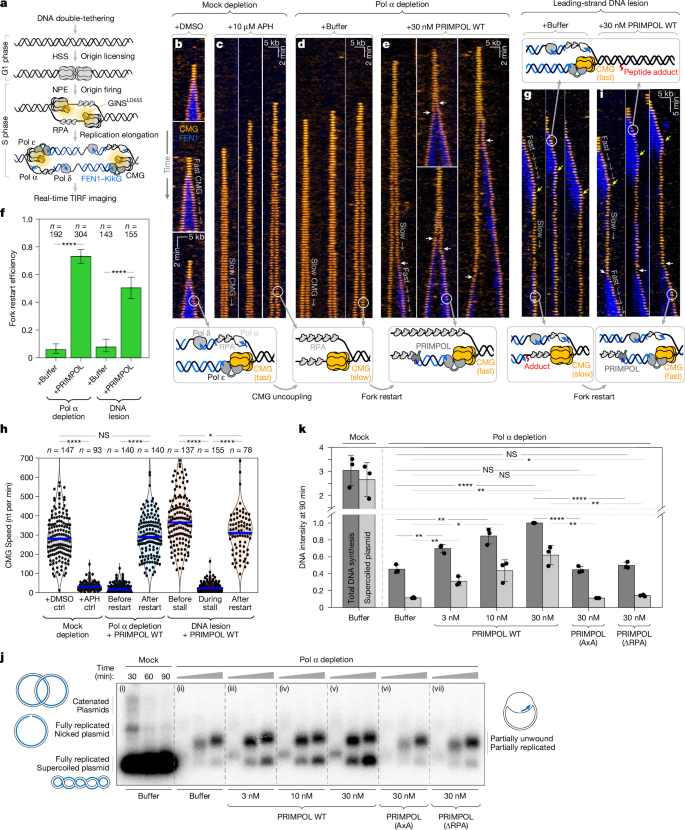
a, The single-molecule imaging workflow. b, Kymograms showing CMG dynamics at unperturbed forks. GINS–LD655 (orange) tracks the CMG helicase, and FEN1–KikG (blue) serves as a nascent DNA marker. c, CMG dynamics in aphidicolin-treated extract. d, CMG dynamics in Pol-α-depleted extract. e, CMG dynamics in Pol-α-depleted extract with 30 nM of WT PRIMPOL. The white arrows denote PRIMPOL-dependent fork restart events. f, Fork restart efficiency: the fraction of uncoupled CMGs that resumed fast translocation. Data are mean ± 95% confidence interval (CI) estimated by bootstrapping. n values denote the number of forks. g, CMG dynamics at DNA lesions in undepleted extract. The yellow arrows indicate fork stalling at the lesion. h, CMG speeds measured in different experiments. The blue lines indicate medians, shaded areas represent 95% CI estimated by bootstrapping. n values denote the number of CMG molecules. Ctrl, control. i, CMG dynamics at DNA lesions in extract + 30 nM of WT PRIMPOL. The white arrows indicate PRIMPOL-mediated restart events. The experiments shown in b–h were repeated twice with different extract preparations. j, Autoradiogram of plasmid replication assay in mock-depleted (i) versus Pol-α-depleted extract + buffer (ii), +3 nM WT PRIMPOL (iii), +10 nM WT PRIMPOL (iv), +30 nM WT PRIMPOL (v), +30 nM PRIMPOL(AxA) (vi) or +30 nM PRIMPOL(ΔRPA) (vii). The cartoons illustrate replication intermediates corresponding to bands on the gel (blue indicates nascent DNA). k, Quantification of the plasmid replication assay shown in j from three independent repeats. Total DNA synthesis (dark grey) or supercoiled DNA (light grey) signal was measured at 90 min and normalized to the total signal of the 30 nM WT PRIMPOL reaction. Data are mean ± s.d. from three independent experiments. P values were computed using two-sample two-sided Kolmogorov–Smirnov tests (f and h) and two-tailed two-sided Student’s t-tests (k); NS, not significant (P > 0.05); P < 0.05, P < 0.01, *P < 0.0001.
To visualize CMG, we used GINS-depleted extract supplemented with fluorescently labelled GINS (GINS–LD655). To monitor DNA synthesis, we supplemented extract with FEN1–KikG (flap endonuclease 1 fused to the fluorescent protein KikG)35. FEN1 is recruited to chromatin through its interaction with PCNA36, and was previously validated as a marker of nascent DNA37, which does not require FEN1’s enzymatic activity37.
In mock-depleted extract, origin firing generated two diverging CMGs travelling at around 300 nucleotides per min with FEN1–KikG signal accumulating behind each helicase (Fig. 1b). To induce RS, the extract was supplemented with 15 µM aphidicolin (APH)—an inhibitor of B-family polymerases (including α, ε and δ) that does not inhibit PRIMPOL (Extended Data Fig. 1d). In APH-treated extract, CMG travelled around 10× slower than in mock without any accompanying FEN1–KikG signal, indicating a failure to synthesize DNA (Fig. 1c). This substantial CMG slowing during RS (reported by us25 and others38) is a hallmark of CMG uncoupling28 (Fig. 1b,c). Adding 30 nM of wild-type (WT) PRIMPOL to APH-treated extract did not support fork restart (Extended Data Fig. 1e), reflecting the fact that APH inhibits Pol ε and δ and prevents them from extending PRIMPOL’s primers.
To recapitulate RS without inhibiting Pol ε/δ, we devised two independent strategies. First, we immunodepleted Pol α from nuclear extract (depleting or inhibiting Pol α was previously used to induce RS in cells22,39,40,41). In Pol-α-depleted extract, CMGs travelled very slowly (around 30 nucleotides per min) without any accompanying FEN1 accumulation (Fig. 1d). This is indicative of helicase uncoupling and is consistent with reports that Pol α is required for establishing leading strand synthesis42,43. When Pol-α-depleted extract was supplemented with recombinant WT PRIMPOL, we routinely observed that slow/uncoupled CMGs suddenly sped up to around 300 nucleotides per min (Fig. 1e (white arrows)). We interpret this switch to fast CMG progression as fork restart. Around 75% of stalled forks restarted in Pol-α-depleted extract supplemented with 30 nM WT PRIMPOL, versus only around 5% restart efficiency in reactions supplemented with buffer (Fig. 1f and Extended Data Fig. 1f). Importantly, the sudden switch from slow to fast CMG progression was accompanied by FEN1–KikG accumulation behind the fork, indicative of DNA synthesis (Fig. 1e). To corroborate the idea that fast CMG progression corresponds to DNA synthesis, we repeated these experiments with fluorescently labelled PCNA (PCNA–LD655)—an established marker of nascent DNA44,45. Like FEN1–KikG, PCNA–LD655 accumulated behind restarted forks (Extended Data Fig. 1g). Disrupting FEN1’s interaction with PCNA eliminated FEN1–KikG binding (Extended Data Fig. 1h) without impacting fork restart (Extended Data Fig. 1i). We conclude that FEN1–KikG is recruited to nascent DNA by PCNA, and that the sudden speed-up of CMG represents fork restart. We subsequently used FEN1–KikG as a DNA synthesis marker because it provided better signal-to-noise than PCNA–LD655 (Methods).
Second, we designed a DNA substrate with site-specific DNA lesions on the leading strand template, each consisting of a small (around 4 amino acids) peptide adduct (Extended Data Fig. 2a). This lesion should easily fit through CMG’s central channel46, and should induce helicase uncoupling. After encountering the lesion, CMG instantly slowed down from around 300 nucleotides per min to 30 nucleotides per min (Fig. 1g,h), indicative of uncoupling. In the absence of PRIMPOL, forks stalled at the lesion restarted very inefficiently (around 7%) (Fig. 1f and Extended Data Fig. 1f), probably through TLS as shown previously47,48. In extract containing 30 nM of WT PRIMPOL, around 50% of forks stalled at the lesion restarted (Fig. 1i,f), causing CMG to speed up from around 30 nucleotides per min to 300 nucleotides per min—matching the speed of unperturbed forks (Fig. 1h and Extended Data Fig. 2b). Importantly, lesion-stalled forks restarted in a PRIMPOL-dependent manner in extract containing endogenous levels of Pol α.
We conclude that recombinant human WT PRIMPOL supports robust fork restart in our single-molecule assay using two orthogonal sources of RS (Fig. 1f). We subsequently used the Pol α depletion method because it provided a higher throughput, and was easily adapted for ensemble assays (see below).
Monitoring PRIMPOL-dependent fork restart
To establish an ensemble assay for PRIMPOL-dependent fork restart, we replicated a 3 kb plasmid in Pol-α-depleted extract, which strongly suppressed DNA synthesis in comparison to the mock-depleted extract (Fig. 1j (compare i and ii)). The low levels of DNA synthesis in Pol-α-depleted extract (Fig. 1j (ii)) are probably caused by trace amounts of Pol α that evaded the immunodepletion (Extended Data Fig. 2c).
Supplementing Pol-α-depleted extract with 3–30 nM of WT PRIMPOL led to a significant increase in DNA synthesis, in a PRIMPOL-concentration-dependent manner (Fig. 1j,k). As human cells contain around 17 nM of PRIMPOL49, the concentrations used here are near physiological. Notably, 30 nM of WT PRIMPOL did not fully rescue DNA synthesis (Fig. 1j (v versus i)). We attribute this to the fact that depleting Pol α activates the DNA damage checkpoint (Extended Data Fig. 2c), which partially inhibits origin firing and results in fewer forks (Extended Data Fig. 2d (CDC45 blot)). We revisit CHK1’s role in Fig. 4.
To verify that PRIMPOL is responsible for the partial rescue of DNA synthesis in the biochemical assay, we tested two previously characterized mutants: (1) PRIMPOL D114A/E116A (hereafter, PRIMPOL(AxA)), which lacks primase and polymerase activity2,3; and (2) PRIMPOL D519R/F522A/D551R/I554A (hereafter, PRIMPOL(ΔRPA)), which cannot interact with RPA30 (Extended Data Fig. 1c). The in vitro primase–polymerase assay showed that PRIMPOL(ΔRPA) (but not PRIMPOL(AxA)) exhibits catalytic activity (Extended Data Fig. 1d). Chromatin fractionation experiments indicate that WT PRIMPOL and PRIMPOL(AxA) (but not PRIMPOL(ΔRPA)) are recruited to DNA in Pol-α-depleted extract (Extended Data Fig. 2d) and in APH-treated extract (Extended Data Fig. 2e). In contrast to WT PRIMPOL, neither PRIMPOL(AxA) nor PRIMPOL(ΔRPA) rescued DNA synthesis in Pol-α-depleted extract (Fig. 1j (v–vii)), corroborating reports that repriming requires both PRIMPOL’s enzymatic activity1,4 and its interaction with RPA30,31. These data indicate that Pol-α-depleted extract recapitulates PRIMPOL-dependent fork restart, providing a powerful model system to dissect the molecular mechanism of repriming.
Long-lived PRIMPOL binding near stalled forks
It was previously proposed that RPA recruits PRIMPOL to stalled forks4,30. As uncoupled CMGs generate long tracts of RPA-coated ssDNA, it remained unclear where exactly PRIMPOL binds along ssDNA. To address this question, we visualized AlexaFluor647-labelled WT PRIMPOL (PRIMPOL(WT)–AF647) in Pol-α-depleted extract supplemented with FEN1–KikG, and RPA fused to mCherry-XL50,51 (hereafter, RPA–mCherryXL) (Extended Data Fig. 3a). This enabled us to simultaneously monitor PRIMPOL binding and dissociation, DNA synthesis and the extent of RPA–ssDNA. Importantly, fluorescent labelling did not impair PRIMPOL’s fork restart activity (Extended Data Fig. 3b,c).
At 30 nM of PRIMPOL(WT)–AF647, there were too many PRIMPOL binding events on RPA–ssDNA, making it impossible to resolve individual molecules (Extended Data Fig. 3d). To distinguish each PRIMPOL binding event, we visualized 3 nM PRIMPOL(WT)–AF647 at 15 s per frame (Fig. 2a). Initially, RPA–ssDNA tracts grew bidirectionally very slowly (Fig. 2a and Extended Data Fig. 3e)—consistent with uncoupled helicases (Fig. 1h). After PRIMPOL-mediated fork restart, RPA–ssDNA tracts grew rapidly, but the RPA signal intensity decreased around twofold (Fig. 2a (bottom)). This suggests that PRIMPOL reprimes synthesis only on one strand (the leading strand, as shown in Fig. 4).
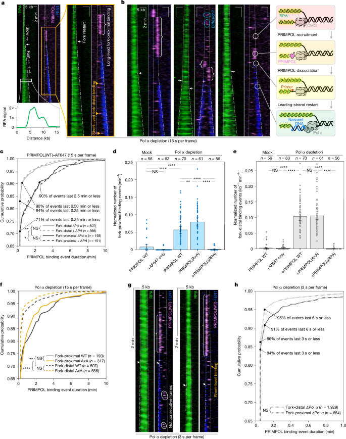
a, Kymogram showing ssDNA coated with RPA–mCherryXL (green) and nascent DNA coated with FEN1–KikG (blue) in Pol-α-depleted extract with 3 nM PRIMPOL(WT)–AF647 (magenta). For clarity, the RPA kymogram is shown separately from FEN1 + PRIMPOL. The grey lines mark the boundary of the RPA signal: the dotted lines denote uncoupled CMGs and the dashed lines mark restarted forks. The white arrows indicate fork restart. Bottom inset: integrated RPA signal profile, which decreases by around 50% after fork restart. b, RPA–mCherryXL (green), FEN1–KikG (blue) and PRIMPOL(WT)–AF647 (magenta) binding to DNA in Pol-α-depleted extract. The cartoons illustrate the fork status over time. c, Cumulative probability plot of PRIMPOL(WT)–AF647 (3 nM) binding event duration, comparing fork-proximal (dark grey) and fork-distal (light grey) binding events in Pol-α-depleted extract (solid lines) or APH-treated extract (dashed lines). n values denote the number of binding events analysed. d, The number of all fork-proximal PRIMPOL binding events per unit time at the fork. e, The number of fork-distal PRIMPOL binding events observed per unit time normalized by the length of exposed ssDNA. In d and e, n values denote the number of forks. Data are mean ± 95% CI, estimated by bootstrapping. f, Cumulative probability plot of fork-proximal (solid lines) and fork-distal (dashed lines) binding events in Pol-α-depleted extract for PRIMPOL(WT)–AF647 (dark grey) and PRIMPOL(AxA)–AF647 (orange). n values denote the number of binding events analysed. Data in a–f are from experiments imaged at 15 s per frame. g, Kymograms showing fork restart in Pol-α-depleted extract with 3 nM PRIMPOL(WT)–AF647 (magenta) imaged at 3 s per frame. h, Cumulative probability plot of PRIMPOL(WT)–AF647 binding event duration in Pol-α-depleted extract imaged at 3 s per frame (at this temporal resolution, short-lived events dominate both fork-distal and fork-proximal subpopulations). n values denote the number of binding events analysed. P values were computed using two-sample two-sided Kolmogorov–Smirnov tests (c–f and h); ***P < 0.001. All experiments were performed twice, with different extract preparations.
We detected numerous short-lived PRIMPOL(WT)–AF647 binding events far from replication forks (Fig. 2a (orange triangles)), which we call fork-distal. Most fork-distal PRIMPOL binding events (~84%) lasted one frame (15 s) or less (Fig. 2c) and were randomly distributed along RPA–ssDNA. We also detected fork-proximal PRIMPOL binding events close to the fork (within 1 pixel = 160 nm), which tended to be longer than fork-distal binding events (Fig. 2c). Notably, long-lived fork-proximal PRIMPOL(WT)–AF647 binding (Fig. 2a,b (curly brackets)) preceded most (~70 ± 10%) fork restart events (Fig. 2a,b (white arrows)).
To gain further insights, we quantified the frequency of fork-proximal (Fig. 2d) and fork-distal (Fig. 2e) binding events in Pol-α-depleted extract supplemented with 3 nM of fluorescently labelled WT PRIMPOL, PRIMPOL(AxA) or PRIMPOL(ΔRPA). As expected, PRIMPOL(ΔRPA) exhibited very little fork-proximal or fork-distal binding in Pol-α-depleted extract, indistinguishable from the negative control, for which we measured the frequency of non-specific binding using free AF647 dye (Fig. 2d,e and Extended Data Fig. 3f). Both WT PRIMPOL and PRIMPOL(AxA) exhibited high levels of fork-proximal and fork-distal binding in Pol-α-depleted extract, consistent with chromatin fractionation experiments (Extended Data Fig. 2d,e). Notably, PRIMPOL(AxA) exhibited the most fork-proximal binding (Fig. 2d), probably because it can repeatedly bind to stalled forks without restarting them (Extended Data Fig. 3g). By contrast, WT PRIMPOL promotes fork restart, after which it no longer binds to the unstressed fork (the mock-depleted control is shown in Fig. 2d and Extended Data Fig. 3h). Notably, there was no statistical difference between binding event durations for WT PRIMPOL and PRIMPOL(AxA) (Fig. 2f), even though only WT PRIMPOL supported fork restart (Extended Data Fig. 2i).
We also observed long-lived fork-proximal WT PRIMPOL binding events before fork restart on the template with a site-specific lesion (Extended Data Fig. 3j). These productive WT PRIMPOL binding events lasted around 3 min, similar to those measured in Pol-α-depleted extract (Extended Data Fig. 3j). Notably, there was a delay of 3–5 min between the dissociation of PRIMPOL(WT)–AF647 from the fork, and fork restart (Extended Data Fig. 3j). This delay probably reflects the time needed for a replicative polymerase to capture the primer synthesized by PRIMPOL, and for the reassembly of an active replisome.
Occasionally, we observed PRIMPOL binding in consecutive movie frames at nearby positions on DNA (Fig. 2b (light blue ovals)). This could represent a PRIMPOL molecule diffusing along ssDNA, or may represent different molecules binding at slightly different positions along ssDNA. To distinguish between these scenarios, we repeated the experiment at 3 s per frame (Fig. 2g, five times faster than in Fig. 2a–f). At this higher frame rate, we frequently observed different PRIMPOL molecules briefly binding to ssDNA in non-consecutive video frames (Fig. 2g (white ovals)). Such binding sequences could be misinterpreted as diffusion of a single PRIMPOL molecule if imaged at lower framerates. This experiment also revealed that the vast majority of PRIMPOL binding events are very short-lived, lasting 3 s or less (Fig. 2h). We still observed long-lived fork-proximal binding events that preceded fork restart (Fig. 2g (white curly brackets)). In summary, although we cannot rule out PRIMPOL diffusion across short distances, our data argue against long-range PRIMPOL diffusion on ssDNA.
Recruitment to stalled forks via interaction with CMG
Although PRIMPOL can transiently bind anywhere along ssDNA–RPA (Fig. 2a,b), the vast majority (over 99%) of binding events are non-productive—that is, they do not lead to fork restart. Productive PRIMPOL binding events occur predominantly near stalled forks and tend to be long-lived (~3 min; Extended Data Fig. 3j), raising the question of why fork-proximal binding events are longer. We hypothesized that PRIMPOL’s binding at the fork may be stabilized by yet unknown interactions with fork-associated proteins. Notably, AlphaFold3 predicted that several replisome subunits may interact with PRIMPOL52. By cross-referencing AlphaFold predictions for human and frog proteins, we singled out PRIMPOL’s putative interaction with MCM4—a CMG subunit (Fig. 3a). Positively charged residues in human PRIMPOL (Arg29, Arg31, Lys34) are predicted to form salt bridges with MCM4 residues (Fig. 3a and Extended Data Fig. 4a,b). To test this prediction, we generated three mutants: PRIMPOL R29A/R31A/K34A (hereafter, PRIMPOL(3A)), PRIMPOL R29D/R31E/K34D (hereafter, PRIMPOL(DED)) and PRIMPOL(29–35:Linker) (in which residues 29–35 were replaced with the linker GGGSGGGS) (Fig. 3b and Extended Data Fig. 4c,d).
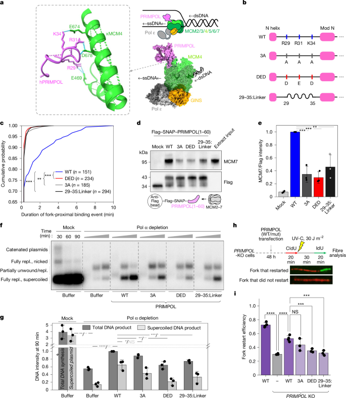
a, View of the predicted interaction between human PRIMPOL residues 28–40 (pink) and Xenopus MCM4 residues 466–479 and 670–689 (green) (left). Right, the PRIMPOL–MCM4 model superimposed onto the cryo-EM structure of the core human replisome (PDB: 7PFO). b, PRIMPOL mutants designed to disrupt the interaction with MCM4. c, The cumulative probability distribution of fork-proximal binding events for PRIMPOL constructs shown in b. n values denote the total number of binding events. d, Representative western blot image from a co-immunoprecipitation (co-IP) experiment in which Flag–SNAP–PRIMPOL(1–60) constructs were immobilized on anti-Flag beads and incubated with Xenopus extract. e, Quantification of the co-IP experiment shown in d from three independent repeats. Data are mean ± s.d. f, Representative autoradiogram of the replication assay in mock-depleted or Pol-α-depleted extract with 30 nM WT PRIMPOL, 30 nM PRIMPOL(3A), 30 nM PRIMPOL(DED) or 30 nM PRIMPOL(29–35:Linker). Repl., replicated. g, Quantification of replication assay shown in f from three independent repeats. Total DNA synthesis (dark grey) and supercoiled plasmid (light grey) signal was measured at 90 min and normalized to the total signal of the WT PRIMPOL control. Data are mean ± s.d. from three independent experiments. h, Diagram of the DNA fibre fork restart assay. PRIMPOL-knockout (KO) cells were transfected with plasmids encoding WT PRIMPOL, PRIMPOL(3A), PRIMPOL(DED) or PRIMPOL(29–35:Linker). Forks were pulse-labelled with CldU (red), irradiated with UV-C, allowed to recover and then pulse-labelled with IdU (green). Representative fibre tracks show a fork that restarted and a fork that did not. i, Quantification of fork restart efficiency measured in DNA fibre experiments (the number of restarted forks divided by the total number of forks). Data are mean ± s.d. from four independent experiments (WT, KO, KO + WT) or three independent experiments (KO + 3A, KO + DED, KO + 29-35:Linker). P values were computed using two-sample two-sided Kolmogorov–Smirnov tests (c) and two-tailed two-sided Student’s t-tests (e, g and i).
First, we measured the duration of fork-proximal PRIMPOL binding events in APH-treated extract. APH does not alter the duration of PRIMPOL-binding events (Fig. 2c) but allows PRIMPOL to repeatedly bind without promoting fork restart (Extended Data Fig. 4e). All three PRIMPOL mutants exhibited significantly reduced fork-proximal binding times compared with WT PRIMPOL (Fig. 3c), suggesting that disrupting the predicted PRIMPOL–MCM4 impairs PRIMPOL recruitment to stalled forks.
Second, we co-immunoprecipitated MCM2–7 from extract using PRIMPOL residues 1–60 as bait. We compared the WT bait versus the 3A, DED and 29-35:linker mutants (Extended Data Fig. 4f). All three mutant bait proteins co-immunoprecipitated with MCM2–7 less efficiently than the WT PRIMPOL(1–60) bait (Fig. 3d,e), corroborating AlphaFold’s prediction.
Third, we examined whether the three full-length PRIMPOL mutants can reprime stalled forks in Pol-α-depleted extract. PRIMPOL(DED) and PRIMPOL(29–35:Linker) mutants exhibited significant defects in DNA synthesis while PRIMPOL(3A) showed a more subtle defect (Fig. 3f,g). Notably, all three mutants exhibited similar enzymatic activity in vitro (Extended Data Fig. 4d), consistent with the fact that residues 29–35 are in a disordered region distant from the catalytic core. Notably, AlphaFold3 predicted that PRIMPOL may also interact with MCM753, which is located next to MCM4 within CMG (Extended Data Fig. 4b). These additional interactions with MCM7 may explain the modest defect of PRIMPOL(3A) in the biochemical fork restart assay (Fig. 3f,g). To disrupt PRIMPOL’s interaction with both MCM4 and MCM7, we prepared the mutant PRIMPOL K8E/E13R/R15E/R29D/R31E/K34D (PRIMPOL(EREDED)). Unfortunately, PRIMPOL(EREDED) exhibited diminished enzymatic activity in vitro (Extended Data Fig. 4d), so we did not investigate it further.
Finally, we examined whether the PRIMPOL–MCM4 interaction is important for fork restart in human cells, using an established DNA fibre assay1 (Fig. 3h). Plasmids encoding WT or mutant PRIMPOL were transiently transfected into PRIMPOL-deficient HCT116 cells. Both PRIMPOL(DED) and PRIMPOL(3A) mutants were expressed at levels similar to WT PRIMPOL, while PRIMPOL(29–35:Linker) was expressed at lower levels that were similar to endogenous PRIMPOL (Extended Data Fig. 4g). To measure the fork-restart efficiency, ongoing forks were pulse-labelled with 5-chloro-2′-deoxyuridine (CldU) for 20 min and cells were irradiated with UV-C light (30 J m−2) to arrest fork progression. Cells were allowed to recover for 30 min and were pulse-labelled with 5-iodo-2′-deoxyuridine (IdU) for 20 min to label only forks that restarted (Fig. 3h). DNA fibre analysis revealed that the PRIMPOL(DED) and PRIMPOL(29–35:Linker) mutants significantly impaired fork restart compared with the WT control (Fig. 3i and Extended Data Fig. 4h). While the PRIMPOL(3A) mutant also impaired fork restart, it exhibited more variability between experimental repeats, rendering the difference between WT PRIMPOL and PRIMPOL(3A) not statistically significant (Fig. 3i). The findings presented in Fig. 3 indicate that the PRIMPOL–MCM4 interaction helps target PRIMPOL to stalled forks, and is required for efficient fork restart.
PRIMPOL also supports CMG-independent restart
We found that WT PRIMPOL promotes efficient fork restart at concentrations as low as 3 nM (Fig. 2a,b). In the low PRIMPOL concentration regime, restart involves long-lived PRIMPOL binding to the fork through MCM4. Consequently, we refer to these events as CMG-dependent restart. Notably, at higher concentrations of WT PRIMPOL (>10 nM), we observed a distinct subpopulation of restart events that occurred far behind the fork (Fig. 4a (white asterisks)). The frequency of these fork-distal restart events increased proportionally to WT PRIMPOL concentration (Fig. 4b (grey bars)), indicating that they also represent repriming.
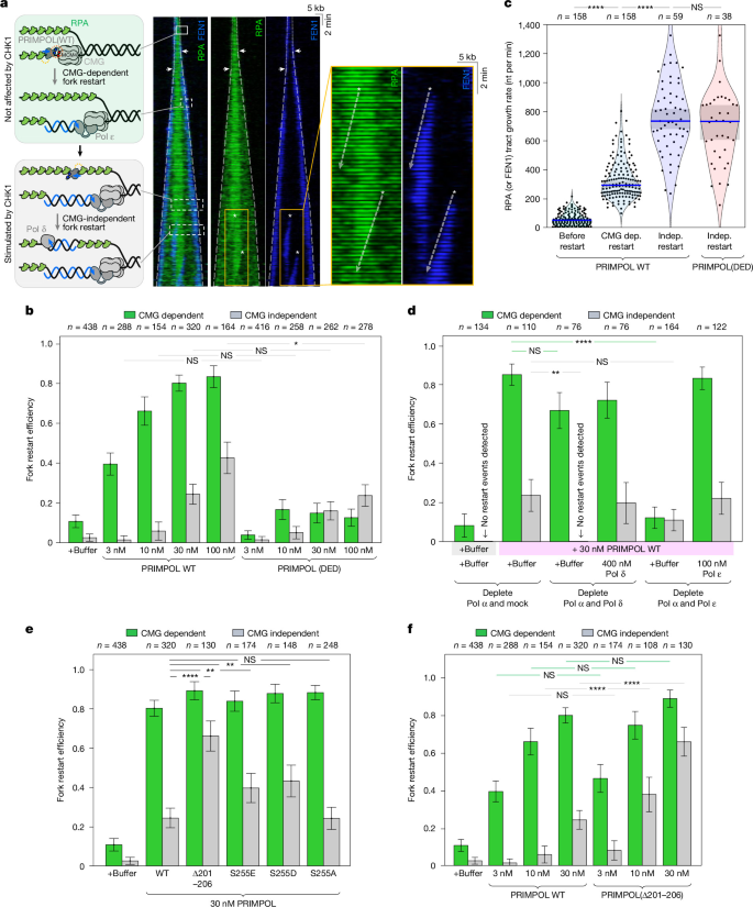
a, Kymogram showing a bidirectional replication bubble undergoing fork restart in Pol-α-depleted extract with 30 nM unlabelled WT PRIMPOL. RPA–mCherryXL (green) and FEN1–KikG (blue) are shown overlaid or separately. The white arrows indicate CMG-dependent fork restart. The white asterisks indicate CMG-independent restart events. Both types of restart event cause RPA clearing from a parental DNA strand. b, CMG-dependent (green) and CMG-independent (grey) restart efficiency in Pol-α-depleted extract supplemented with buffer, or various concentrations of WT PRIMPOL or PRIMPOL(DED). c, DNA synthesis rates measured in Pol-α-depleted extract supplemented with 30 nM of unlabelled WT PRIMPOL or PRIMPOL(DED) extracted from RPA tract growth (before and after CMG-dependent restart) or from FEN1 tract growth (after CMG-independent (indep.) restart). The blue lines indicate the median values and the shaded boxes represent the 95% CI estimated by bootstrapping. n values denote the number of restart events. d, Fork restart efficiency measured in Pol-α- and mock-depleted extract; Pol-α- and Pol-δ-depleted extract; and Pol-α- and Pol-ε-depleted extract. The reactions were supplemented with 30 nM of unlabelled WT PRIMPOL and 400 nM Pol δ or 100 nM Pol ε. e, The fork restart efficiency measured in Pol-α-depleted extract supplemented with 30 nM of unlabelled WT PRIMPOL or PRIMPOL phosphomutants (PRIMPOL(Δ201–260), PRIMPOL(S255E), PRIMPOL(S255D), PRIMPOL(S255A)). f, The fork restart efficiency measured in Pol-α-depleted extract supplemented with increasing concentrations of WT PRIMPOL or PRIMPOL(Δ201–260). For b–f, data are mean ± 95% CI, estimated by bootstrapping, n values denote the number of forks. P values were computed using two-sample two-sided Kolmogorov–Smirnov tests. All experiments were performed twice with different extract preparations.
We repeated experiments with PRIMPOL(DED) and observed a substantial decrease in CMG-dependent restarts (Fig. 4b (green bars)), but not in fork-distal restarts (Fig. 4b (grey bars) and Extended Data Fig. 5a,b). This suggests that the latter represent a distinct type of restart event, which we call CMG independent. Notably, the FEN1-labelled nascent DNA tracts grew significantly faster during CMG-independent restart (~700 nucleotides per min) than during CMG-dependent restart (~300 nucleotides per min) (Fig. 4c and Extended Data Fig. 5c), corroborating our hypothesis that CMG-dependent and CMG-independent restarts are mechanistically distinct (see below).
We conclude that PRIMPOL supports two distinct types of restart event: (1) CMG-dependent restart that requires the PRIMPOL–CMG interaction and is efficient at limiting PRIMPOL concentrations; and (2) CMG-independent restart that does not require the PRIMPOL–CMG interaction and becomes efficient only at high PRIMPOL concentrations that mimic overexpression15.
Pol ε is required for CMG-dependent fork restart
The PRIMPOL–CMG interaction raises the possibility that PRIMPOL’s primer could be handed directly to the leading strand polymerase Pol ε, which is physically tethered to CMG54,55,56 in a manner that should not prevent PRIMPOL from binding to MCM4 (Fig. 3a). If so, Pol ε could capture PRIMPOL’s primer and extend it to restart leading strand synthesis. Alternatively, PRIMPOL’s primer could be extended by Pol δ first, and then handed to Pol ε, reminiscent of how leading strand synthesis is established during replication initiation42,57.
To test these hypotheses, we performed experiments in extract depleted of Pol α and Pol δ, or in extract depleted of Pol α and Pol ε (Fig. 4d). Depleting Pol δ caused only a minor reduction in CMG-dependent fork restart efficiency, whereas depleting Pol ε ablated CMG-dependent restart events (Fig. 4d (green bars) and Extended Data Fig. 6a–c). Importantly, CMG-dependent fork restart was fully rescued by adding 100 nM (Pol ε concentration in human cells49) of recombinant Pol ε to Pol-ε-depleted extract (Fig. 4d and Extended Data Fig. 6d). These data support the hypothesis that CMG-dependent restart entails the primer handover from PRIMPOL to Pol ε, which subsequently performs long-range DNA synthesis.
Notably, depleting Pol δ (but not Pol ε) eliminated CMG-independent restart events (Fig. 4d (grey bars) and Extended Data Fig. 6a). Supplementing Pol-δ-depleted extract with 400 nM (Pol δ concentration in human cells)49 of recombinant Pol δ fully rescued CMG-independent restart (Fig. 4d and Extended Data Fig. 6e,f). These findings suggest that CMG-independent restart entails the primer handover from PRIMPOL to Pol δ, which subsequently performs long-range DNA synthesis.
We conclude that PRIMPOL primarily acts as a leading-strand primase by synthesizing primers immediately behind CMG, and those primers are extended by Pol ε. However, at higher concentrations, PRIMPOL can also reprime DNA synthesis in a CMG-independent manner, and those primers are extended by Pol δ. Our observation that DNA synthesis proceeds faster during CMG-independent restart (Fig. 4c) may reflect the fact that this process does not require template unwinding, or may point to Pol δ being able to synthesize DNA faster than Pol ε.
CHK1 activation is not required for repriming
It was previously reported that CHK1 phosphorylates human PRIMPOL at Ser255, which promotes more repriming and RS tolerance in human cells22. The same study proposed that a disordered loop in human PRIMPOL (residues 201–260) autoinhibits repriming, but that Ser255 phosphorylation by CHK1 relieves this autoinhibition22. We examined whether CHK1 modulates PRIMPOL’s activity in our biochemical fork restart assay. We supplemented Pol-α-depleted extract with an ATR inhibitor (AZD6738) that blocked CHK1 phosphorylation (Extended Data Fig. 7a (lane iii versus iv)). Notably, inhibiting ATR in Pol-α-depleted extract enhanced the amount of PRIMPOL-dependent DNA synthesis (Extended Data Fig. 7b (v versus vi) and 7c). This indicates that checkpoint activation is not required for PRIMPOL to restart stalled forks. This finding also explains the low levels of DNA synthesis reported in Fig. 1j: depleting Pol α activates the DNA damage checkpoint, which suppresses origin firing, resulting in fewer replication forks (Extended Data Fig. 2d (CDC45 immunoblot)). Thus, although PRIMPOL can restart most stalled forks in Pol-α-depleted extract, the overall amount of DNA synthesis is expected to be lower than in the mock control (Fig. 1j (i versus v)).
Immunoblotting of phosphorylated CHK1 in Pol-α-depleted extract collected from the flow cell revealed that the checkpoint is not active in our single-molecule experiments. This is because the DNA template concentration in the flow-cell is very low (10−3−10−2 ng μl−1) while checkpoint activation requires at least around 1 ng μl−1 (ref. 58) of DNA template. The fact that we observe robust PRIMPOL-dependent fork restart in the single-molecule assay (Fig. 1f) in the absence of CHK1 activation (Extended Data Fig. 7a (flow-cell lane)) supports our conclusion that checkpoint activation is not required for repriming.
CHK1 upregulates CMG-independent repriming
Given the report that CHK1 stimulates PRIMPOL activity in human cells22, we hypothesized that CHK1 may regulate another aspect of PRIMPOL’s function. To test this idea, we performed single-molecule fork restart experiments with four previously characterized PRIMPOL phospho-mutants22 (Extended Data Fig. 7d,e): phospho-dead PRIMPOL(S255A), phosphomimetic PRIMPOL(S255E) and PRIMPOL(S255D), and a mutant that phenocopies the phosphomimetic mutants—PRIMPOL(Δ201–260) (in which residues 201–260 were replaced by a flexible linker). At a near-physiological concentration (30 nM), all four mutants supported similar levels (80–90%) of CMG-dependent fork restart to WT PRIMPOL (Fig. 4e (green bars) and Extended Data Fig. 7f). However, the mutants supported different levels of CMG-independent restart (Fig. 4e (grey bars)): PRIMPOL WT and PRIMPOL(S255A) exhibited the lowest efficiency (~25%); PRIMPOL(S255E) and PRIMPOL(S255D) supported intermediate efficiency (~40%); while PRIMPOL(Δ201–260) displayed the highest efficiency (~70%).
We further examined PRIMPOL(Δ201–260) across a wide range of concentrations (Fig. 4f). At near-physiological concentrations (10 nM and 30 nM), PRIMPOL(Δ201–260) supported significantly more CMG-independent restart than WT PRIMPOL (Fig. 4f (grey bars) and Extended Data Fig. 7g). We conclude that CHK1 phosphorylation of Ser255 does not affect PRIMPOL’s primary mode of repriming (CMG dependent), but significantly enhances PRIMPOL’s secondary repriming mode (CMG independent). Consistent with our conclusion, the stimulatory effect of CHK1 phosphorylation on PRIMPOL’s activity was previously observed in cells treated with a Pol α inhibitor22—which exposes large tracts of ssDNA behind the fork, generating the ideal substrate for CMG-independent repriming.
Discussion
Our experiments revealed several mechanistic insights into PRIMPOL function, dynamics and regulation, supporting a new model of PRIMPOL-mediated fork restart (Fig. 5). First, PRIMPOL can briefly bind anywhere along RPA–ssDNA (Fig. 5a–c), but such binding events are very short-lived (Fig. 5c) and rarely cause fork restart. Second, PRIMPOL can reside on RPA–ssDNA much longer near uncoupled CMGs (Fig. 5d–f), and such long-lived binding events frequently lead to fork restart. Third, the PRIMPOL–MCM4 interaction is required for efficient fork restart (Fig. 5e) in nuclear extract and in human cells. Finally, our data indicate that, at near-physiological concentrations, PRIMPOL primarily acts as a leading-strand primase, and that PRIMPOL’s primers are directly handed to Pol ε (Fig. 5f–h). Notably, PRIMPOL-driven restart can also occur far behind stalled forks (Fig. 5i–l), but only at high PRIMPOL concentrations (mimicking overexpression), or after ATR/CHK1 activation. It is tempting to speculate that chemotherapy-induced PRIMPOL overexpression observed in cancer cells14,15 may unlock this alternative CMG-independent restart mechanism. As PRIMPOL cannot prime on ssDNA fully coated with RPA30, CMG-independent repriming events may occur only when a RPA molecule transiently dissociates from ssDNA in PRIMPOL’s immediate vicinity (Fig. 5i,j). Notably, RPA can spontaneously dissociate from ssDNA in vitro59, albeit slowly (koff = 0.006 s−1).
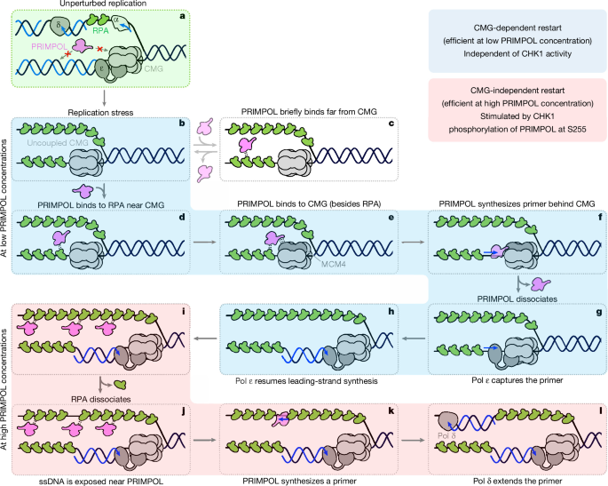
a, During unperturbed replication, Pol ε and Pol δ synthesize the leading and lagging strands, respectively, minimizing the accumulation of RPA-coated ssDNA. b, During RS, CMG becomes uncoupled from DNA synthesis and continues to unwind DNA, generating RPA-coated ssDNA. c, PRIMPOL transiently binds to RPA far from CMG, but such fork-distal binding events are short-lived and rarely lead to repriming. d, PRIMPOL binds to RPA near CMG. e, PRIMPOL simultaneously interacts with both RPA and MCM4, which prolongs PRIMPOL’s residence at the stalled fork. f, Long-lived fork-proximal binding enables PRIMPOL to synthesize a short primer behind CMG. g, After PRIMPOL dissociates, Pol ε captures the primer synthesized by PRIMPOL. h, Pol ε elongates the primer and restarts leading-strand synthesis. i, At high PRIMPOL concentrations, there are many short-lived PRIMPOL binding events on RPA–ssDNA. j, Infrequent RPA dissociation generates exposed ssDNA near PRIMPOL. k, PRIMPOL synthesizes a primer far behind CMG. l, Pol δ captures the primer synthesized by PRIMPOL and elongates it, restarting DNA synthesis independently of CMG. b–h, (blue-shaded panels) CMG-dependent fork restart is efficient at low PRIMPOL concentrations and occurs independently of CHK1 signalling. i–l, (red-shaded panels) CMG-independent fork restart becomes more frequent at higher PRIMPOL concentrations (which mimic overexpression) and depends on CHK1 phosphorylation of PRIMPOL at Ser255.
We show that efficient repriming requires both PRIMPOL–RPA and PRIMPOL–MCM4 interactions. As RPA binds to PRIMPOL’s C terminus30 and MCM4 binds to PRIMPOL’s N terminus (Fig. 3), we propose that PRIMPOL simultaneously interacts with both RPA and MCM4 during repriming (Fig. 5e). This model provides simple-yet-elegant answers to three long-standing questions. First, PRIMPOL can interact with both RPA and MCM4 only behind uncoupled CMGs, explaining how PRIMPOL is targeted specifically to stalled forks (Fig. 5e). Second, the simultaneous interaction with both RPA and MCM4 positions PRIMPOL near CMG’s exit channel (Fig. 3a). This enables PRIMPOL to engage the leading-strand template before it is coated by RPA (Fig. 5f), explaining how repriming can occur in the nucleus where RPA is very abundant. Third, PRIMPOL’s binding to CMG facilitates rapid primer handover to Pol ε (Fig. 5g), minimizing error-prone DNA synthesis during repriming. It is unclear what causes PRIMPOL’s dissociation after primer synthesis but before fork restart (Fig. 2b). It is possible that PRIMPOL disengages RPA once the primer becomes too long (Fig. 5f), which could promote PRIMPOL’s dissociation from DNA. Alternatively, PRIMPOL may transiently dissociate from MCM4 while CMG recruits replication factors that prevent PRIMPOL from re-binding the helicase.
Notably, we observed a negligible amount of PRIMPOL binding to unperturbed forks (mock in Fig. 2d,e and Extended Data Fig. 3h). This could be because, during normal replication, there is no RPA-coated ssDNA immediately behind CMG (Fig. 5a). Alternatively, during unperturbed replication, PRIMPOL’s docking site on CMG may be sterically occluded by a replication elongation factor. For example, CTF18 binds to the C terminus of MCM7 (ref. 45) (next to MCM4), which may prevent PRIMPOL binding to CMG during replication elongation.
During the preparation of this manuscript, we became aware of a similar study60 from the Yeeles group (MRC Laboratory of Molecular Biology, Cambridge, UK), which reconstituted PRIMPOL-dependent fork restart using purified human proteins and obtained a cryo-electron microscopy (cryo-EM) structure of the CMG–PRIMPOL complex that is highly similar to the AlphaFold prediction (personal communication).
There are a few notable similarities between PRIMPOL’s role in the nucleus and mitochondria. First, PRIMPOL interacts with the mitochondrial replicative helicase Twinkle61 and the ssDNA-binding protein mtSSB9, reminiscent of its interactions with CMG and RPA. Notably, PRIMPOL also interacts with the mitochondrial DNA polymerase γ61, but there is no evidence of PRIMPOL interacting with Pol ε/δ. Second, PRIMPOL is required to restart replication after mitochondrial DNA (mtDNA) damage61, similar to its role in restarting forks in the nucleus. Notably, Twinkle can stimulate PRIMPOL’s enzymatic activity in vitro62, but it remains unclear how this regulates repriming in mitochondria. Third, PRIMPOL overexpression leads to increased levels of lagging strand priming during mtDNA replication, reminiscent of CMG-independent restart events observed by us at high PRIMPOL concentrations (Fig. 4b (grey bars)). The ability of PRIMPOL to restart DNA synthesis far behind CMG may reflect a vestigial or primordial role of PRIMPOL to prime mtDNA lagging strand synthesis. This activity is normally suppressed by ssDNA-binding proteins (RPA or mtSSB), but becomes possible after PRIMPOL overexpression. Notably, DNA damage checkpoint activation and the subsequent phosphorylation of PRIMPOL by CHK1 (ref. 22) further upregulate CMG-independent repriming (Fig. 4e,f).
Although PRIMPOL is regarded as an attractive therapeutic target17,63, currently there are no small-molecule PRIMPOL inhibitors. Our findings suggest an alternative strategy to inhibit repriming—namely, to prevent PRIMPOL binding to CMG. This could be accomplished using peptides or molecules that bind to PRIMPOL’s N terminus, which normally interacts with MCM4–CMG.
Methods
X. laevis
Eggs from adult laboratory-bred female X. laevis frogs (Xenopus 1, 4800) were used to prepare egg extracts. Testes from adult laboratory-bred male X. laevis frogs (Xenopus 1, 4293) were used to prepare sperm chromatin. All animals were healthy, 2–3 years of age, not subjected to previous procedures and no animal husbandry was performed. No animal randomization or blinding was performed. Frogs were housed at the Boswell Small Amphibians Research Facility at Stanford School of Medicine and all experiments involving frogs were approved by Stanford’s Institute Animal Care and Use Committee (IACUC-33670).
Human cell lines
PRIMPOL-proficient and PRIMPOL-KO HCT116 cells were generated in the Méndez laboratory and cultured in Dulbecco’s modified Eagle’s medium (Sigma-Aldrich, D5796) supplemented with 10% FBS (Biowest S1810) plus 1% penicillin–streptomycin (Solmeglas, L0018). All human cell lines were routinely tested for mycoplasma contamination and consistently tested negative throughout the course of the study.
Insect cell lines
Sf9 cells (Expression Systems, 94-001S) and Tni cells (Expression Systems, 94-002S) were cultured in ESF 921 insect cell culture medium (Expression Systems, 96-001-01) at 27 °C for baculovirus production and protein overexpression.
Bacterial cell lines
DH10EMBacY (Geneva Biotech, DH10EMBacY), DH5α (New England Biorabs (NEB), C2987H) and Rosetta (DE3) pLysS (Novagen, 70956-4) bacteria were cultured in TB (RPI Research Products, T15050) or LB broth (Thermo Fisher Scientific, BP9723) at 37 °C for plasmid production and protein overexpression.
IVTT system
In vitro transcription and translation (IVTT) reactions were performed using the TnT SP6 High-Yield Wheat Germ Protein Expression System (Promega, L3261) according to the manufacturer’s instructions. Proteins were not purified after production by IVTT.
Xenopus egg extract preparation
X. laevis egg extracts were prepared according to published protocols64,65. Frogs were primed with 50 IU of human chorionic gonadotropin (CHORULON) (Merck, 133754) 5 days before extract preparation. To induce ovulation, 500 IU of CHORULON was injected 21–22 h before extract preparation. Sperm chromatin was purified from adult male frog testes and used for nucleoplasmic extract (NPE) preparation.
Cloning and plasmids
To prepare antigens consisting of X. laevis PRIMPOL amino acids 1–212, X. laevis POLA1 amino acids 1–340, POLD1 amino acids 1–89, POLE1 amino acids 578–754 and full-length X. laevis PCNA, the corresponding DNA sequence was PCR amplified out of X. laevis egg cDNA. The resulting PCR product was assembled into pET28b(+) using the NEBuilder HiFi Mix (NEB, E2621S), yielding plasmids pJP001 (PRIMPOL), pGC279 (POLA1), pLDL048 (POLD1), pLDL037 (POLE1) and pLDL190 (PCNA).
To prepare human WT His6-SNAP-PRIMPOL, synthetic gene blocks were ordered from Integrated DNA Technologies (IDT) and assembled into pET28b(+) using the NEBuilder HiFi Mix (NEB, E2621S), yielding plasmid pJP012.
To prepare the PRIMPOL(AxA) mutant, a gene block containing D114A/E116A was ordered from IDT and assembled into a PCR-amplified vector backbone using primers oJP100 and oJP101 with pJP012 as the template, yielding plasmid pJP057.
To prepare the PRIMPOL(ΔRPA) mutant, a gene block containing D519R/F522A/D551R/I554A was ordered from IDT and assembled into a PCR-amplified vector backbone using primers oJP098 and oJP102 with pJP012 as the template, yielding plasmid pJP058.
To prepare the PRIMPOL(3A), PRIMPOL(DED) and PRIMPOL(29–35:Linker) mutants, gene blocks containing R29A/R31A/K34A; R29D/R31E/K34D; and 29–35aa to GGGSGGGS, respectively, were ordered from IDT and assembled into a PCR-amplified vector backbone using primers oJP083 and oJP84 with pJP012 as the template, yielding plasmids pJP093, pJP052 and pJP051, respectively.
To prepare the PRIMPOL(Δ201–260) mutant, gene blocks containing the 201–260aa to SGSETPGTSESATPES mutation was ordered from IDT and assembled into a PCR-amplified vector backbone using primers oJP007 and oJP008 with pJP012 as the template, yielding plasmid pJP013. To prepare the PRIMPOL(S255E), PRIMPOL(S255D) and PRIMPOL(S255A) mutants, primers oJP022 (for S255E), oJP023 (for S255D) and oJP024 (for S255A) were ordered from IDT and PCR-amplified with oJP021 using pJP012 as the template. PCR products were then phosphorylated (NEB, M0201) and ligated (NEB, M0202), yielding plasmid pJP102, pJP103 and pJP104, respectively.
To prepare human WT His6–Flag–SNAP–PRIMPOL1–60, pJP012 was PCR-amplified using primers oJP098 and oJP114. The PCR product was resolved by native agarose gel electrophoresis and purified using NucleoSpin Gel and PCR Clean-up kit (Macherey-Nagel, 740609) according to the manufacturer’s instructions. Purified DNA was phosphorylated with T4 polynucleotide kinase (NEB, M0201) and ligated using T4 DNA Ligase (NEB, M0202), yielding plasmid pJP084.
To prepare human mutant His6–Flag–SNAP–PRIMPOL1-60 variants (3A, DED and 29-35:Linker), the same cloning strategy described above was used, yielding plasmid pJP095, pJP085 and pJP097, respectively.
Detailed procedures for cloning the plasmid encoding full-length X. laevis Pol δ are described in our recent publication66.
Plasmids containing the individual subunits of X. laevis Pol ε with sequences codon optimized for insect cell expression were a gift from J. Walter. The individual subunits were cloned from the following vectors. The pole1 gene was assembled with a 3×Flag-3aalinker-hrv3c-s6-10aalinker containing geneblock (synthesized by IDT) into pACEBac1, yielding plasmid pLDL114. The pole2 gene was assembled into pIDC, yielding pLDL055. The pole3 gene was assembled into pIDC, yielding pLDL056. The pole4 gene was assembled into pIDC, yielding pLDL057. The three small subunits were then cloned into a single plasmid using the MultiBac multigene construction strategy67. The pole3 gene was extracted from pLDL056 and inserted into linearized pLDL057, yielding pLDL058. The pole2 gene was extracted from pLDL055 and inserted into linearized pLDL058, yielding pLDL059. The pLDL114 and pLDL059 vectors were fused by Cre-recombination, yielding pLDL132. The 3×Flag tag was replaced by a TwinStrep tag by Gibson assembly, yielding the complete insect expression plasmid pLDL158.
To prepare X. laevis His6-5aalinker-s6-10aalinker-PCNA cloning, synthetic gene blocks were ordered from IDT and assembled into pET28b(+) using the NEBuilder HiFi Mix (NEB, E2621S), yielding plasmid pJP087. Detailed information on the primer oligonucleotides used in this study is listed in Supplementary Table 2.
Recombinant protein expression, purification
Previously published protocols were used to purify recombinant GINS25. FEN1–KikG, RPA–mCherry, RPA–mKikGR, human SNAP–PRIMPOL (WT and mutants) and X. laevis s6–PCNA were purified using the following protocols. Rosetta (DE3) pLysS cells overexpressing each protein were collected and the cell pellets were resuspended in lysis buffer (500 mM NaCl, 50 mM HEPES pH 7.5, 30 mM imidazole, 10% glycerol, 5 mM β-mercaptoethanol and 1 mM PMSF). Cells were lysed by sonication, and the lysates were cleared by ultracentrifugation at 35,000 rpm for 1 h at 4 °C using the Beckman Type 70 Ti rotor. The cleared lysates were incubated with 0.5 ml of Ni-NTA resin for 1 h at 4 °C. Bound protein was eluted with elution buffer identical to the lysis buffer except containing 400 mM imidazole.
For FEN1–KikG, the imidazole eluate was sufficiently pure and was therefore dialysed into storage buffer (150 mM NaCl, 50 mM HEPES pH 7.5, 1 mM dithiothreitol (DTT) and 10% glycerol), flash-frozen and stored at −80 °C. s6–PCNA was further purified by FPLC using the HiTrap Heparin HP affinity column (Cytiva, 17040701), followed by a Superdex 200 Increase 10/300 GL column (Cytiva, 28990944). The Ni–NTA eluate containing 500 mM NaCl was diluted with buffer A (20 mM HEPES pH 7.5, 0.5 mM EDTA, 1 mM DTT, and 10% glycerol) to a final salt concentration of 150 mM and loaded onto the heparin column. Bound proteins were eluted over a 150–750 mM KCl gradient using a mixture of buffer A and buffer B (buffer A supplemented with 1 M KCl). The fractions corresponding to the protein peak (typically eluting at ~30% buffer B, ~300 mM KCl) were collected and concentrated for subsequent size-exclusion chromatography. Heparin-purified proteins were further purified and buffer-exchanged into storage buffer (150 mM NaCl, 50 mM HEPES pH 7.5, 1 mM DTT and 10% glycerol) by size-exclusion chromatography on the Superdex 200 Increase 10/300 GL column. Peak fractions were pooled, concentrated (Amicon Ultra Centrifugal Filter, 3 kDa MWCO, Millipore, UFC800324), flash-frozen and stored at −80 °C. For other proteins, the Ni-NTA eluate was further purified by FPLC using a HiTrap Heparin HP affinity column (Cytiva, 17040701), followed by a Superdex 200 Increase 10/300 GL column (Cytiva, 28990944). The Ni–NTA eluate containing 500 mM NaCl was diluted with buffer A (20 mM HEPES pH 7.5, 0.5 mM EDTA, 1 mM DTT and 10% glycerol) to a final salt concentration of 150 mM and loaded onto the heparin column. Bound proteins were eluted over a 150–750 mM KCl gradient using a mixture of buffer A and buffer B (buffer A supplemented with 1 M KCl). Fractions corresponding to the protein peak (typically eluting at ~30% buffer B, ~300 mM KCl) were collected and concentrated for subsequent size-exclusion chromatography. Heparin-purified proteins were further purified and buffer-exchanged into storage buffer (150 mM NaCl, 50 mM HEPES pH 7.5, 1 mM DTT and 10% glycerol) by size-exclusion chromatography on the Superdex 200 Increase 10/300 GL column. The peak fractions were pooled, concentrated (Amicon Ultra Centrifugal Filter, 3 kDa MWCO, Millipore, UFC800324), flash-frozen and stored at −80 °C. For s6–PCNA, the HiTrap Q HP affinity column (Cytiva, 17115301) was used, followed by the Superose 6 Increase 10/300 GL column (Cytiva, 29091596) after fluorescent labelling. The buffer conditions were the same.
Flag–SNAP–PRIMPOL(1–60) proteins (WT and mutants) used for co-IP assays were purified exactly same as described above using the same buffers. The Ni-NTA eluate was further purified by FPLC using a HiTrap Heparin HP affinity column using the same procedure. The fractions corresponding to the Flag–SNAP–PRIMPOL(1–60) peak were collected and concentrated, flash-frozen and stored at −80 °C.
Full-length X. laevis Pol δ was purified as follows. A bacmid was generated by transforming DH10EMBacY cells with [pLDL158] pBiG_TwinStrep-HRV3C-S6-Pol δ, and the resulting bacmid was purified using the ZR BAC DNA Miniprep Kit (Zymo Research, D4049). Baculovirus was amplified in three 72 h stages (P1, P2, P3) in Sf9 cells. We initially attempted to express Pol δ in Tni Cells; however, the POLD3 subunit was not recovered (presumably due to proteolysis). Instead, TwinStrep-HRV3C-S6-Pol δ was expressed in 2 l of Sf9 suspension culture by infection with 4 ml of P3 baculovirus for 65 h. Cells were collected, pelleted by centrifugation and flash-frozen in liquid nitrogen. Cell pellets were thawed and resuspended in 200 ml of lysis buffer (25 mM HEPES-NaOH pH 7.5, 300 mM NaCl, 10% glycerol, 1 mM DTT—bubbled with argon gas to remove dissolved oxygen) supplemented with 1× Pierce Protease Inhibitor EDTA-free (Thermo Fisher Scientific, A32965) and lysed by dounce homogenization. The lysates were cleared by ultracentrifugation at 35,000 rpm for 45 min at 4 °C in a Beckman Type 70 Ti rotor (Beckman Coulter). Cleared lysates were incubated with 1 ml of Strep-Tactin XT 4 Flow resin (IBA Lifesciences, 2-5010-010) for 1 h at 4 °C. The resin was washed three times with 10 ml lysis buffer. The protein was eluted from the resin using 4 ml buffer N supplemented with 50 µg ml−1 HRV3C protease for 2 h at 4 °C on a rotator, and flash-frozen in liquid nitrogen. The eluent was thawed and then diluted with 2 ml of no-salt buffer (25 mM HEPES-NaOH pH 7.5, 10% glycerol, 1 mM DTT) to reduce the NaCl concentration to 200 mM, and loaded onto the Mono Q 5/50 GL column (Cytiva, 17516601). The protein was eluted in 20 column volumes (CVs) from the MonoQ column with a linear gradient of 200 mM–1,000 mM NaCl and eluted around 350 mM NaCl. The peak fractions were pooled, spin-concentrated to about 0.5 ml using the Amicon Ultra-4-10K column (Merck Millipore, UFC801024) and loaded onto a Superdex 200 Increase 10/300 gel-filtration column (Cytiva, 28-9909-44) equilibrated in 25 mM HEPES-NaOH pH 7.5, 10% glycerol, 1 mM DTT and 150 mM NaCl. The peak fractions were pooled, spin-concentrated to about 4 µM using the Amicon Ultra-4-10K column (Merck Millipore, UFC801024), and frozen in liquid nitrogen and stored at −80 °C. Approximately 50 µg total protein was obtained from 2 l of Sf9 cells.
Full-length X. laevis Pol ε was purified as follows. A bacmid was generated by transforming DH10EMBacY cells with [pLDL158] pBiG_TwinStrep-HRV3C-S6-Pol ε, and the resulting bacmid was purified using the ZR BAC DNA Miniprep Kit (Zymo Research, D4049). Baculovirus was amplified in three 72 h stages (P1, P2, P3) in Sf9 cells. TwinStrep-HRV3C-S6-Pol ε was expressed in 1 l of Tni suspension culture by infection with 2 ml of P3 baculovirus for 48 h. Cells were collected, pelleted by centrifugation and flash-frozen in liquid nitrogen. Cell pellets were thawed and resuspended in 100 ml of buffer N (25 mM HEPES-NaOH pH 7.5, 300 mM NaCl, 10% glycerol, 1 mM DTT, bubbled with argon gas to remove dissolved oxygen) supplemented with 1× Pierce Protease Inhibitor EDTA-free (Thermo Fisher Scientific, A32965) and lysed by dounce homogenization. The lysates were cleared by ultracentrifugation at 35,000 rpm for 45 min at 4 °C in a Beckman Type 70 Ti rotor (Beckman Coulter). The cleared lysates were then incubated with 1 ml of Strep-Tactin XT 4 Flow resin (IBA Lifesciences, 2-5010-010) for 1 h at 4 °C. The resin was washed three times with 10 ml buffer. The protein was eluted from the resin using 4 ml buffer N supplemented with 50 μg ml−1 HRV3C protease for 2 h at 4 °C on a rotator, and flash-frozen in liquid nitrogen. Eluent was thawed and diluted with buffer A (25 mM HEPES-NaOH pH 7.5, 10% glycerol, 1 mM DTT) to reduce the NaCl concentration to 200 mM, and loaded onto the Mono Q 5/50 GL column (Cytiva, 17516601). The protein was eluted in 20 CVs from the MonoQ column with a linear gradient of 200 mM–1,000 mM NaCl in buffer A, and eluted at around 350 mM NaCl. The peak fractions were pooled, spin-concentrated to around 0.5 ml using the Amicon Ultra-4-10K column (Merck Millipore, UFC801024) and loaded onto a Superdex 200 Increase 10/300 gel-filtration column (Cytiva, 28-9909-44) equilibrated in 25 mM HEPES-NaOH pH 7.5, 10% glycerol, 1 mM DTT and 150 mM NaCl. The peak fractions were pooled, spin-concentrated to about 4 μM using the Amicon Ultra-4-10K column (Merck Millipore, UFC801024) and frozen in liquid nitrogen and stored at −80 °C.
The denaturing purification of antigens was performed as follows. The Rosetta (DE3) pLysS cell pellet overexpressing His6-tagged antigen was thawed in denaturing lysis buffer (50 mM HEPES pH 7.5, 500 mM NaCl, 30 mM imidazole, 6 M urea) and sonicated. The lysates were cleared by ultracentrifugation at 35,000 rpm for 1 h at room temperature in the Beckman Type 70 Ti rotor. The cleared lysates were then incubated with 0.5 ml of Ni-NTA resin for 1 h at room temperature. The protein was eluted with 400 mM imidazole in denaturing elution buffer (50 mM HEPES pH 7.5, 500 mM NaCl, 6 M urea).
Antibody preparation
Custom polyclonal antibodies were generated as previously described34. Most home-made antibodies used in this study were raised by Cocalico Biologicals using a protocol approved by the Institute Animal Care and Use Committee (IACUC-33724). Antibodies were affinity purified from sera in the lab, except for Xenopus PRIMPOL antibody 2 (amino acids 662–674; L3754/3755), which was raised and affinity-purified by Biosynth. All antibodies used in this study were affinity purified. In total, 1–5 mg of purified antigen was cross-linked to 1 ml of AminoLink Plus Coupling Resin (Thermo Fisher Scientific, PI20501) according to the manufacturer’s protocol. Then, 10–30 ml of serum was incubated with the affinity resin for 30 min at room temperature. The resin was washed twice with 10 ml of 1× PBS + 500 mM NaCl, and twice again with 10 ml of 1× PBS. Next, antibodies bound to the affinity resin were eluted 5–6 times with 1.0 ml of 200 mM glycine-HCl pH 2.5. Each elution was collected into a tube containing 0.1 ml of 1 M Tris-HCl pH 9.0, which immediately neutralized the glycine. Eluted fractions were pooled and dialysed against 1 l of 1× PBS + 10% (w/v) sucrose. The IgG concentration was measured using the Thermo Fisher Scientific NanoDrop UltraC (Thermo Fisher Scientific), normalized to 1 mg ml−1 through dilution or spin-concentration, aliquots were flash-frozen in liquid nitrogen and stored at −80 °C. Human PRIMPOL monoclonal antibodies (mouse MAb 106E and rat MAb 71D), were raised in the Méndez laboratory and the CNIO monoclonal antibody facility13. All commercially available antibodies were validated by the manufacturers. Non-commercially available antibodies were generated and validated by the indicated laboratories. For example, antibodies used for western blotting were validated using the corresponding recombinant proteins as positive controls. Antibodies used for immunodepletion were validated by IP followed by western blot analysis of the supernatant and bead-bound fractions. The specificity of immunodepletion was further assessed by add-back of the corresponding recombinant protein to the immunodepleted extract. Detailed information on the antibodies used in this study is provided in Supplementary Table 3.
Preparation of DNA substrates
For ensemble DNA replication assays, 3 kb pBlueScript II SK(+) plasmid DNA was purified using the QIAprep Spin Miniprep Kit (Qiagen, 27106) and used as the replication templates. All single-molecule experiments used doubly tethered λ phage DNA, except for the experiments shown in Fig. 1h,i and Extended Data Fig. 2d, which used DNA templates containing two site-specific leading strand lesions (small peptide adducts). The detailed protocol for the preparation of biotinylated λ DNA is described in our recent methods paper34. Preparation of DNA templates containing two site-specific leading strand lesions (DNA–protein cross-links, DPCs) has been described previously25. To generate small peptide adducts rather than bulky DPCs, DPC-containing DNA templates were treated with 1 µL of 120 U ml−1 thermolabile proteinase K (NEB, P8111) at room temperature for 15 min, followed by heat inactivation at 55 °C for 10 min before use.
Single-molecule replication assay
All single-molecule experiments were conducted according to a previously published protocol34 at temporal resolutions of 3, 15, 30 or 60 s per frame. During the λ DNA tethering step, 20 pM of Streptavidin–AF647 (Thermo Fisher Scientific, S21374) was added to the streptavidin solution. Streptavidin–AF647 spots were used as a fiducial marker for focusing immediately before starting imaging. After λ DNA tethering, DNA was licensed in undepleted high-speed supernatant (HSS) containing 26 ng µl−1 of 30 bp carrier oligonucleotide and ATP regeneration system (ARS) for 3 min. After licensing, DNA was washed with 1× ELB-sucrose, and a replication mix containing of 33% NPE, 33% HSS, 20 ng µl−1 pBlueScript carrier plasmid DNA, ARS, and unlabelled or fluorescently labelled proteins (typically, 20 nM GINS–LD655, 200 or 830 nM FEN1–KikG, 300 nM RPA–mCherry, 3–100 nM PRIMPOL or PRIMPOL–AF647, 100 nM Pol ε, 400 nM Pol δ and 300 nM PCNA–LD655) was flowed into the microfluidic chamber. When using Pol-α-depleted extract, 10 µM of CD437 was also added to the reaction mixture to suppress the activity of trace amounts of endogenous Pol α that evaded the immunodepletion (we used the ensemble plasmid replication assay to verify that this concentration of CD437 does not impair PRIMPOL’s activity).
The OpenCell database estimates that human cells contain around 17 nM of PRIMPOL49. However, a substantial portion of PRIMPOL is in mitochondria, and there are no reliable estimates of PRIMPOL’s concentration in the nucleus. Moreover, the exact concentration of nuclear PRIMPOL is likely to vary between cell types. For this reason, we sampled a wide range of PRIMPOL concentrations (3–100 nM), but we refer to concentrations of 10–30 nM as near-physiological based on OpenCell estimates.
To visualize fluorescently labelled PCNA–LD655 during fork restart (Extended Data Fig. 1g,h), we immunodepleted >95% of endogenous PCNA from extract and supplemented this depleted extract with 500 nM of PCNA–LD655. We estimate that NPE and HSS contain ~30 µM and ~1 µM of PCNA, respectively, corresponding to ~10 µM PCNA in the final replication reaction (consisting of 33% NPE, 33% HSS and 33% buffer). After immunodepletion, around 300 nM of endogenous PCNA remained in the final reaction mixture (ensemble plasmid replication assay confirmed that this residual PCNA concentration was sufficient to support normal DNA replication). Supplementation with 300 nM PCNA–LD655 therefore resulted in an approximately 1:1 ratio of fluorescently labelled to endogenous PCNA in the experiments shown in Extended Data Fig. 1g,h. However, imaging fluorescently labelled PCNA in this system is technically challenging because PCNA is highly abundant in egg extracts and these experiments require simultaneous immunodepletion of Pol α (to induce RS) and PCNA (to reduce the concentration of endogenous/unlabelled PCNA). Without PCNA depletion, addition of 300 nM PCNA–LD655 to extracts containing ~10 µM endogenous PCNA in the final reaction would result in only a small fraction of the total PCNA being fluorescently labelled, substantially reducing the signal-to-noise ratio. For this reason, we used PCNA–LD655 imaging as an orthogonal validation of FEN1–KikG as a marker of nascent DNA rather than as the primary marker of DNA synthesis throughout this study.
GINS–LD655 was imaged using the following settings: total internal reflection fluorescence (TIRF) arm set to 9,000 (~67°), 1 s exposure, 10% 640 nm laser power (~150 µW power out of the objective, ~1.5 W cm−2 power density), 150 EM gain. PRIMPOL–AF647 was imaged using the following settings: TIRF arm set to 9,000 (~67°), 500 ms exposure, 25% 640 nm laser power (~380 µW power out of the objective, ~3.8 W cm−2 power density), 150 EM gain. FEN1–KikG was imaged using the following settings: TIRF arm set to 9,000 (~67°), 500 ms exposure, 8% 488 nm laser power (~70 µW power out of the objective, ~0.7 W cm−2 power density), 150 EM gain. RPAmCherry was imaged using the following settings: TIRF arm set to 9,000 (~67°), 1 s exposure, 10% 561 nm laser power (~70 µW power out of the objective, ~0.7 W cm−2 power density), 150 EM gain. PCNA–LD655 was imaged using the following settings: TIRF arm set to 9,000 (~67°), 1 s exposure, 20% 640 nm laser power (~300 µW power out of the objective, ~3.0 W cm−2 power density), 150 EM gain. Given these exposure durations, 4–10 fields of view (FOVs) could be sequentially imaged during the 15–60 s per frame. A suite of MATLAB scripts written in house was used to select and analyse data as described in our recent methods article34. Below we summarize the data analysis pipeline specific for the single-molecule experiments analysed in this study.
Ensemble replication assay
Ensemble DNA replication experiments were performed following a detailed protocol described in a recent methods article34. In brief, licensing mixtures contained mock- or Pol-α-depleted HSS, ARS, pBlueScript plasmid DNA and α-32P-dCTP. Replication reactions were initiated by addition of mock- or Pol-α-depleted NPE supplemented with ARS and PRIMPOL proteins (WT or mutants). We chose to use human PRIMPOL because: (1) PRIMPOL is highly conserved across vertebrates2; (2) we could easily interrogate mutants that were previously characterized in human PRIMPOL; and (3) human PRIMPOL expresses well in bacteria3. Reactions were quenched at the indicated timepoints by addition of 1× stop buffer (10 mM Tris pH 8.0, 1% SDS, 30 mM EDTA, 10% glycerol and 0.1% (w/v) bromophenol blue) containing proteinase K (1 mg ml−1 final concentration). The samples were analysed by 0.8% native agarose gel electrophoresis. After electrophoresis, gels were dried, exposed to a phosphor screen and analysed by phosphor imaging using the Azure Sapphire Gel Imaging System (Azure Biosystems).
Western blotting and Coomassie staining
All of the samples were boiled in 1× SDS–PAGE sample buffer (50 mM Tris-HCl pH 6.8, 2% SDS, 10% glycerol and 0.05% (w/v) bromophenol blue) and run on 4–15% Mini-PROTEAN TGX Precast Protein Gels (Bio-Rad, 4561086) using Tris-glycine-SDS running buffer (Boston BioProducts BP-150). Colour Prestained Protein Standard Broad Range (NEB, P7719) was run alongside samples to infer protein band sizes. For Coomassie staining, protein gels were incubated with Optiblot Blue InstantBlue Coomassie Protein Stain (Abcam, ab119211) for at least 1 h at room temperature with gentle shaking. For immunoblotting, proteins were transferred from the gel onto a PVDF membrane (VWR, 10061-492) in transfer buffer (Boston BioProducts, BM190). Membranes were blocked in 1× PBST (1× PBS (Boston BioProducts, BM220), 0.05% Tween-20) containing 5% (w/v) non-fat milk for 1 h at room temperature with gentle shaking. After a wash in 1× PBST, the membrane was incubated with primary antibodies diluted in 1× PBST containing 1% (w/v) BSA overnight at 4 °C with gentle shaking. The membranes were washed three times with 1× PBST and incubated with secondary antibody diluted in 1× PBST + 5% non-fat milk for 1 h at room temperature with gentle shaking. For immunoblotting of Ser345-phosphorylated CHK1, 1× TBST (1× Tris-buffered saline (TBS) Boston BioProducts, BM301, 0.05% Tween-20 for immunoblotting) was used instead of PBST and the membrane was blocked in 1× TBST containing 10% (w/v) BSA. The membranes were washed three times with 1× PBST (or TBST), developed using SuperSignal West Pico PLUS Chemiluminescent Substrate (Thermo Fisher Scientific, PI34577) and imaged using ChemiDoc MP Imaging System (Bio-Rad).
In vitro PRIMPOL primase–polymerase assay
The in vitro PRIMPOL primase–polymerase assay was performed according to a previously published protocol68. In brief, a 5′-T36CCTGT20-3′ ssDNA was used as the template. The reactions were carried out in buffer containing 50 mM HEPES pH 7.5, 1 mM MnCl2, 10 μM dNTP and SYBR Green I at a dilution of 1:20,000. Reaction mixtures contained 30 nM ssDNA template and PRIMPOL (WT and mutants) at concentrations ranging from 1 nM to 100 nM. Reactions were incubated in Corning 96-well plates (Corning, 3904) for 1 h at room temperature with moderate agitation. Fluorescence was measured using SpectraMax M2 Microplate Readers (Molecular Devices) with excitation at 497 nm and emission at 520 nm.
Chromatin fractionation assay
For the chromatin fractionation assay, sperm chromatin was used at a final concentration of 1,000 sperm per µl in the reaction mixture. Sperm chromatin was licensed with mock- or Pol-α-depleted HSS (or undepleted HSS for experiments using APH as a source of RS) for 30 min at room temperature. Replication reactions were initiated by addition of mock- or Pol-α-depleted NPE (or DMSO- or APH-treated NPE for APH experiments) supplemented with recombinant PRIMPOL (WT or mutants), followed by incubation for 45 min at room temperature. The reactions were quenched by addition of ice-cold 1× ELB-sucrose (0.25 M) containing 0.2% Triton X-100 and immediately layered onto a 1× ELB-sucrose (0.5 M) cushion. The samples were centrifuged at 15,000g for 1 min at 4 °C to pellet chromatin. The supernatants were carefully aspirated, leaving 15 µl above the pellet. Chromatin pellets were washed twice with ice-cold 1× ELB-sucrose (0.25 M). After washing, chromatin pellets were resuspended in SDS loading buffer containing β-mercaptoethanol and boiled. Samples were resolved by SDS–PAGE, transferred to PVDF membranes and analysed by western blotting using the indicated primary antibodies.
IP for IP–MS assay
IP for MS analysis was performed using Protein A Dynabeads (Invitrogen, 10002D). Dynabeads were incubated with the indicated antibodies (mock control or Xenopus PRIMPOL antibodies 1 or 2) and washed with PBS containing 0.02% Tween-20 to remove unbound antibody. Clarified HSS extract was added to the antibody-bound beads and incubated for 1 h at room temperature with rotation to allow IP. After incubation, beads were washed with ELB-sucrose buffer containing 0.02% NP-40 and subsequently washed with PBS.
MS sample preparation
Samples were trypsin-digested on magnetic beads. The embedded proteins were exchanged into 200 μl of 100 mM triethyl ammonium bicarbonate and reduced with 500 mM of DTT, added to a final concentration of 10 mM. After incubation for 5 min at 55 °C, the beads were mixed head-over-head at room temperature for 25 min on the Barnstead Thermolyne Labquake rotisserie shaker. They were then alkylated with 1 M acrylamide (30 mM final concentration) at room temperature for 30 min. The reduced and alkylated proteins were digested overnight at 37 °C, using 500 ng of MS-grade trypsin/LysC mix (Promega). After digestion, the samples were quenched with formic acid (adjusted to a pH of around 3), eluted from the beads and desalted using the MonoSpin C18 Solid-Phase Extraction (SPE) columns (GL Sciences). Finally, the samples were dried in a SpeedVac SPD 130 DLX vacuum concentrator (Thermo Fisher Scientific) and exchanged into liquid chromatography (LC)–MS reconstitution buffer (2% acetonitrile with 0.1% formic acid in water) for instrumental analysis.
LC–MS/MS analysis
Proteolytically digested peptides were separated using an in-house pulled and packed reverse-phase analytical column (~25 cm length, 100 μm inner diameter), with Dr. Maisch 1.9 μm C18 beads as the stationary phase. Separation was performed with an 80 min reverse-phase gradient (2–45% B, followed by a high-B wash) on the Acquity M-Class UPLC system (Waters) at a flow rate of 300 nl min−1. Mobile phase A was 0.2% formic acid in water, while mobile phase B was 0.2% formic acid in acetonitrile. Ions were formed by electrospray ionization and analysed on an Orbitrap Exploris 480 mass spectrometer (Thermo Fisher Scientific), operated in a data-dependent mode using HCD fragmentation.
LC–MS/MS data analysis
The .raw data were analysed using Byonic v.5.1.1 (Protein Metrics) to identify peptides and infer proteins. A concatenated FASTA file containing UniProt X. Laevis proteins, bait sequences and other likely contaminants and impurities was used to generate an in silico peptide library. Proteolysis with trypsin/LysC was assumed to be fully specific. The precursor and fragment ion tolerances were set to 12 ppm. Cysteine alkylated with propionamide was set as a fixed modification in the search. Variable modifications included oxidation on methionine, dioxidation on tryptophan, N-terminal acetylation, and cyclization of glutamine and glutamic acid. Proteins were held to a false-discovery rate of 1% using the standard reverse-decoy technique69.
Preparation of Xenopus adult tissue cell lysates
Xenopus adult tissues were dissected on ice and washed thoroughly with ice-cold 1× PBS. Tissues collected from female animals included heart, liver, spleen and lung. Ice-cold RIPA lysis buffer supplemented with protease inhibitor was added at a ratio of 50 mg tissue per ml of lysis buffer. Tissues were finely minced using scissors and incubated at 4 °C for 1 h with vigorous vortexing every 10 min. After lysis, the samples were centrifuged at 16,000g for 20 min at 4 °C. The clarified supernatants were collected, and total protein concentrations were determined using a BCA assay. Equal amounts of protein were resolved by SDS–PAGE and analysed by western blotting using a Xenopus PRIMPOL antibody.
Fluorescent labelling of PRIMPOL using SNAP
Labelling of purified human His6–SNAP–PRIMPOL was performed using SNAP-Surface Alexa Fluor 647 (NEB, S9136S). A 1.5-fold molar excess of dye relative to the protein was added and incubated overnight at 4 °C with gentle end-over-end rotation. The reaction mixture was aliquoted, flash-frozen in liquid nitrogen and stored at −80 °C. Labelled PRIMPOL was used directly in single-molecule assays without further purification.
Fluorescent labelling of PCNA using SFP synthase
Purified X. laevis His6–s6–PCNA was fluorescently labelled using in-house purified SFP synthase (surfactin phosphopantetheinyl transferase) and LD655–CoA (purchased from Lumidyne Technologies). S6–PCNA, SFP synthase and LD655–CoA were mixed at a molar ratio of 1:2:10 and incubated at room temperature for 1 h in reaction buffer containing 25 mM HEPES pH 7.5, 60 mM NaCl, 10 mM MgCl2, 1 mM DTT and 10% glycerol. The reaction mixture was further purified using the FPLC-Superose 6 Increase 10/300 GL column (Cytiva, 29091596) to separate unreacted fluorophore and SFP from PCNA–LD655. The peak fractions containing PCNA–LD655 were pooled and concentrated.
Protein co-IP assay
Protein co-IP assays were performed using anti-Flag M2 Magnetic Beads (Millipore, M8823). Flag-tagged SNAP–hPRIMPOL(1–60) proteins (WT or mutants) were purified as described above and immobilized on Flag beads before incubation with Xenopus egg extracts. For each reaction, 3 µl of beads (binding capacity of around 20 pmol Flag peptide) was incubated with 200 pmol of Flag–SNAP–PRIMPOL(1–60) protein (tenfold molar excess relative to bead capacity). Xenopus egg extracts were prepared by mixing NPE and HSS at a 1:1 ratio and diluting fivefold in 1× ELB-sucrose buffer. Beads were then incubated with Xenopus extract for 1 h at room temperature with gentle rotation to allow co-IP of interacting proteins. After incubation, beads were washed with 1× ELB-sucrose containing 0.05% NP-40. Bound proteins were eluted by resuspending beads in SDS sample buffer and boiled at 98 °C for 5 min. Beads were pelleted by brief centrifugation, and only the supernatants were collected for analysis. Eluted proteins and input samples were resolved by SDS–PAGE and analysed by western blotting with the indicated antibodies.
Fork restart assay using stretched DNA fibres
To measure fork restart after replicative stress, cells were first pulsed with 50 μM 5-chloro-2′-deoxyuridine (CldU; Sigma-Aldrich, C6891) for 20 min, irradiated with UV-C (30 J m−2) and, after a 30 min recovery time, pulse-labelled with 250 μM 5-iodo-2′-deoxyuridine (IdU; Sigma-Aldrich, I7125) for 20 min. Cells were collected in ice-cold PBS, resuspended at 0.3 × 10⁶ cells per ml and lysed on microscope slides with spreading buffer (0.2 M Tris pH 7.4, 50 mM EDTA, 0.5% SDS; 6 min at room temperature). DNA fibre spreads were prepared by tilting the slides 15° after cell lysis. The slides were air-dried and fixed in cold methanol:acetic acid (3:1) for 2 min, and incubated in 3 M HCl (30 min at room temperature) to denature DNA. CldU and IdU tracks were detected by immunofluorescence using anti-BrdU monoclonal antibodies with different specificity (rat monoclonal antibody, Abcam, ab6326, for CldU; BD Biosciences mouse monoclonal antibody, 347580, for IdU). The DNA fibre integrity was assessed using a mouse monoclonal anti-ssDNA antibody (DSHB, AB10805144); all incubations were for 1 h at room temperature in a humidity chamber. Standard secondary antibodies were used (anti-mouse IgG1 AF-488, Invitrogen, A21121; anti-rat IgG AF-594, Invitrogen, A11007; anti-mouse IgG2a AF-647, Invitrogen, A21241; all incubations were 30 min at room temperature). The slides were mounted with ProLong Gold antifade reagent (Invitrogen, P36930). Images were acquired on the DM6000B Leica microscope with an HCX PL APO ×40/0.75 NA objective and analysed with ImageJ. For functional complementation assays, HCT116 PRIMPOL-KO cells were transfected with pcDNA3.1/V5-PRIMPOL plasmids expressing the WT form or the indicated mutant derivative (DED, 3A and 29-35:Linker), using Lipofectamine 2000. n = 4 replicates (WT, PRIMPOL KO, PRIMPOL KO + WT) or n = 3 replicates (PRIMPOL KO + DED, PRIMPOL KO + 3A, PRIMPOL KO + 29-35:Linker). 500 replicative structures were counted in each condition and replicate.
Software used
Nikon Elements AR (v.5.20.02) was used to acquire raw microscopy data. ImageJ Fiji (v.2.1.0) was used to stabilize the raw microscopy videos and to quantify signals from autoradiographs and western blots. MATLAB (R2023b) and Origin (2023b) were used to analyse the data. All figures (including cartoons) were created and laid out in Microsoft PowerPoint for Mac (v.16.113.2).
Data reporting
Details of the statistical analyses performed are indicated in the figure legends. As some quantities measured in this study did not have symmetric distributions, we reported the median of the distribution. Unless otherwise specified in the figure captions, error bars represent the 95% CIs for the median. This was calculated using MATLAB’s built in bootstrapping function. The exact meaning of the number ‘n’ reported in each figure is indicated in the figure captions, it usually refers to the number of the number of replication forks or the number of binding events. For single-molecule experiments, we typically used n ≈ 100 events to report. No statistical methods were used to pre-determine sample size. No data blinding or randomization was used. P values were computed using the two-sample two-tailed Kolmogorov–Smirnov test function built into MATLAB, and all experiments were repeated at least twice, with independent extract preparations. For the ensemble DNA replication assay, co-IP assays and cell-based fibre assays, we carried out 3 or 4 independent replicates. P values were computed using the two-sample two-tailed Student t-test function built into MATLAB. The data presented in the Figures are derived from one of the replicates. The exact P values for all data presented in the Figures and the Extended Data Figures are listed in Supplementary Table 4.
Single-molecule data analysis
Each replication initiation experiment imaged 4–10 FOVs over the course of 60–90 min with a resolution of 15, 30 or 60 s per frame. Each FOV typically contained 50–200 DNA molecules stretched to 80–90% of their contour length. A suite of MATLAB scripts written in house were used to select and analyse data as described in our recent methods article70. Below we summarize the data analysis pipeline specific for the single-molecule experiments analysed in this study.
Selecting DNA molecules
Regions of interest corresponding to individual DNA molecules were selected manually based on the previews of DNA stained with SYTOX Orange (these were acquired before the start of the experiment). Only well-separated DNA molecules were used in the analysis.
Fork-speed analysis
To measure fork speed before/after the PRIMPOL-dependent fork restart, we fit the GINS (Fig. 1) or RPA (Fig. 2 and Fig. 4) signals with a linear model (mock + buffer, mock + 10 µM APH, ∆Polα + buffer conditions), a bilinear model (∆Pol α + PRIMPOL, leading-strand DNA lesion + buffer) or a trilinear model (leading-strand DNA lesion + PRIMPOL) in MATLAB and extracted the slope of each linear fit (this has units of pixels per frame). To calculate the fork speed, we multiplied the slope of each linear fit segment to 160 nm per pixel (the size of our camera’s pixel), and divided it by 0.34 nm per bp (to convert nanometres into base pairs), and further divided it by 0.85 (to account for the fact that the DNA template is stretched to around 85% of its contour length).
PRIMPOL-mediated CMG-independent fork restart analysis
In Fig. 4, PRIMPOL-mediated CMG-independent fork restart was monitored by FEN1–KikG signal. To measure the speed of DNA synthesis during CMG-independent restart, the raw FEN1 signal was automatically thresholded using OTSU’s method using a built-in MATLAB function. The OTSU threshold binary image was used to fit straight lines to the edges of the growing FEN1 signal, and the fork speed value was extracted from the slope of the line (accounting for pixel size, the time resolution of the experiment and the extent to which DNA was stretched). Note that the efficiency of CMG-dependent and CMG-independent restart can add up to more than 1. This is illustrated in Fig. 4a, which contains two replication forks: both forks restarted in a CMG-dependent manner (white arrows), and there were two additional CMG-independent restart events (white asterisks).
Analysing PRIMPOL binding-dissociation (Δt 1) and dissociation-fork restart (Δt 2)
To analyse the binding and dissociation (Δt1) of PRIMPOL–AF647, we used the generalized likelihood ratio test (GLRT) as described in our previously study71. Specifically, we used GLRT to convert single-molecule images into binary images, which were subsequently segmented into binding events. The GLRT algorithm accounts for the intrinsic properties of noise in each experiment—a sliding window (we used a five-pixel window) is used to determine whether the signal at any pixel represents noise or signal. Using GLRT, we determined the time of PRIMPOL binding and PRIMPOL dissociation (and therefore Δt1) before a fork-restart event. To calculate the time delay between PRIMPOL–AF647 dissociation and fork restart (Δt2), we first measured the time of PRIMPOL–AF647 dissociation (as described above for Δt1), and the time of fork restart. The time of fork restart was extracted from the bilinear fit to the edge of the RPA signal (Fig. 2a): fork restart corresponds to the changepoint when the fork suddenly speeds up.
PRIMPOL fork-distal versus fork-proximal binding analysis
To measure the location and duration of PRIMPOL–AF647 binding events, we used the GLRT method as described above. To determine whether any given PRIMPOL–AF647 binding event is fork-proximal or fork distal, we compared the position of the binding event along the DNA molecule with the position of the replication fork measured by fitting the fluorescent RPA tract (as illustrated in Fig. 2a). Any PRIMPOL–AF647 binding event within 1 pixel of the replication fork was classified as fork-proximal, and all other binding events were classified as fork-distal.
To quantify the frequency of fork-proximal binding events across different conditions (no RS and RS), or for different PRIMPOL constructs (WT, AxA mutant or RPA-interaction mutant), we measured (1) Nproximal, the number of fork-proximal PRIMPOL–AF647 binding events for each fork; and (2) Tfork, the total duration that we observed that replication fork. We divided Nproximal by Tfork to obtain the normalized quantity shown in Fig. 2d.
We chose a different normalization strategy for fork-distal PRIMPOL–AF647 binding events because they can occur anywhere along RPA-coated ssDNA. To this end, for each replication fork we measured (1) Ndistal, the number of fork-distal binding events; (2) LRPA–ssDNA, the extent of the RPA-coated ssDNA; and (3) Tfork, the total duration of time that the fork was monitored. We divided Ndistal by LRPA–ssDNA and Tfork to obtain the normalized quantity shown in Fig. 2e.
Materials availability
All newly generated reagents generated in this study are available on request from the corresponding author.
Reporting summary
Further information on research design is available in the Nature Portfolio Reporting Summary linked to this article.