 Newshot
Main
Aberrant enhancer-driven transcription sustains many cancers, including metastatic castration-resistant prostate cancer (mCRPC)3,4. AR, a central driver of mCRPC, undergoes ligand-induced activation and dimerization upon binding to androgen response element (ARE)-enriched enhancers and assembling cancer-specific neo-enhanceosomes4,5,6. These complexes depend on the paralogous acetyltransferases p300 and CBP7. p300 has been identified as the predominant paralogue supporting AR-positive (AR+) prostate cancer growth, and the p300/CBP-dependent enhancer mark H2B N-terminal acetylation (H2BNTac) and AR–p300 co-bound enhancer activity are increased in prostate tumours8. Previous studies showed that the N-terminal domain (NTD) of AR interacts with p3002, and this interaction stimulates the histone acetyltransferase (HAT) activity of p300, leading to enhancer remodelling and transcriptional activation by establishing a permissive chromatin environment9,10. Although p300/CBP-targeted therapies have been developed for castration-resistant prostate cancer (CRPC)11,12, global p300/CBP inhibition may cause toxicity in normal tissues13.
Chemically induced proximity (CIP) reprograms protein–protein interactions to manipulate cellular functions14,15. Established CIP modalities include proteolysis-targeting chimeras (PROTACs), which promote target protein degradation via E3 ligase recruitment16; regulated induced proximity targeting chimeras (RIPTACs), which couple disease-associated proteins to essential effectors17,18; transcriptional/epigenetic chemical inducers of proximity (TCIPs), which redirect transcriptional coactivators19,20,21; and targeted relocalization activating molecules (TRAMs), which alter protein localization22. These approaches predominantly connect non-native partners. Here we introduce DALTACs, which enforce neomorphic domain interactions between physiological partners to disable endogenous oncogenic complexes. DALTAC-1 (JZY3032) connects the ligand-binding domain (LBD) of AR to the bromodomain (BRD) of p300/CBP, reprogramming substrate engagement, eliminating H2BNTac and suppressing AR–p300/CBP-dependent transcription. DALTAC-1 outperforms combined AR and p300/CBP inhibition, selectively targets AR-dependent prostate cancer, and induces tumour regression without overt toxicity, establishing domain-level rewiring as a promising therapeutic strategy.
Development of AR–p300/CBP DALTACs
Previous studies established that AR physically interacts with the coactivator p300 through its NTD2, and that dimerized transcription factors can stimulate p300 HAT activity9. To reprogram this native interaction, we designed heterobifunctional molecules that tether an AR-LBD binder to a p300/CBP-BRD binder through an appropriate linker. We reasoned that these molecules would induce non-native AR–p300/CBP interactions and thereby inhibit the growth of AR+ cancer cells by concurrently inhibiting both AR and p300/CBP functions (Fig. 1a).
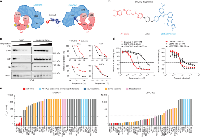
a, Schematic showing how DALTAC-1 rewires domain–domain interactions of the native AR–p300/CBP complex. Created in BioRender; Young, E. https://BioRender.com/ehpy9qp (2026). b, Structure of DALTAC-1 (JZY3032), comprising AR and p300/CBP binders connected by a linker. c, Cellular thermal shift assay of VCaP cells treated with DMSO or DALTAC-1 (100 nM, 4 h). Left, immunoblots showing stabilization of p300, CBP and AR, but not BRD4. Right, protein levels normalized to the lowest temperature condition (three independent experiments). d, Dose–response curves and IC50 values of AR+ VCaP and LNCaP cells treated with DALTAC-1, CBPD-409 or p300/CBPi plus ARi for five days. Data are mean ± s.d.; n = 3 replicated wells; three independent experiments. e, Ranked IC50 values for DALTAC-1 and CBPD-409 across different human cell lines after five-day treatments. Colours indicate specific cell types. PCa, prostate cancer.
To test this premise, we designed and synthesized a series of heterobifunctional molecules using a potent p300/CBP-BRD inhibitor (GNE-04912) and a selective AR-LBD binder23 tethered through a linker (Extended Data Fig. 1a). We selected the tethering site in the p300/CBP-BRD inhibitor based upon the co-crystal structure with GNE-049 and its analogues, which showed that our selected tethering site was exposed to the solvent environment24. Similarly, we chose the tethering site in the AR binder on the basis of our modelled complex structure with AR and extensive structure–activity relationships reported for this class of AR inhibitors23,25. We evaluated our synthesized compounds with proliferation assays in AR+ LNCaP and 22Rv1 cells, which were shown to be responsive to p300/CBP-BRD inhibitors and PROTAC degraders11,26. GNE-049, the AR binder JZY3221, and their combination were included as controls. Treatment with GNE-049, JZY3221 or their combination showed limited inhibitory activity, particularly in 22Rv1 cells (Extended Data Fig. 1b).
To optimize these heterobifunctional molecules, we replaced the tetrahydropyran group of GNE-049 with a piperidinyl group that preserves p300 binding24 and systematically varied linker composition, length and rigidity (Extended Data Fig. 1b and Supplementary Note 1). Several compounds showed substantially greater anti-proliferative activity than the parent-inhibitor combination. Compound 7 (JZY3031) emerged as a lead, with half-maximal inhibitory concentration (IC50) values of 1.9 nM and 2.6 nM in LNCaP and 22Rv1 cells, respectively, and was more potent than the inhibitor combination (Extended Data Fig. 1b).
Replacing an oxygen atom in the AR-binding moiety of compound 7 with an N-methyl group, a modification previously shown to improve oral bioavailability25, generated JZY3032 (Fig. 1b). JZY3032 achieved IC50 values of 0.26 nM and 1.0 nM in LNCaP and 22Rv1 cells, respectively, and was more potent than the parent-inhibitor combination (Extended Data Fig. 1b). JZY3032 also showed improved systemic exposure and 18.5% oral bioavailability in mice, despite its high molecular mass of 975 Da (Extended Data Fig. 1c).
In an enzyme-linked immunosorbent assay (ELISA) measuring induced AR–p300 complex formation, compound 7 and JZY3032 were the most potent and effective compounds that we tested. JZY3032 also produced the strongest inhibition of AR transcriptional activity in HEK293 ARE–GFP reporter cells (Extended Data Fig. 1b). By contrast, three enzalutamide-based bifunctional molecules were substantially less active in VCaP, LNCaP and 22Rv1 cells, demonstrating the importance of the AR-binding moiety (Extended Data Fig. 1d,e).
Based on its cellular, biochemical and pharmacokinetic properties, we designated JZY3032 as DALTAC-1. We also generated Neg-1, with reduced p300/CBP binding24, and Neg-2, with reduced AR binding27 (Extended Data Fig. 1f). Fluorescence polarization assays confirmed their intended binding profiles (Extended Data Fig. 1g–i). Cellular thermal shift assays further showed that DALTAC-1 stabilized AR, p300 and CBP, but not BRD4, supporting selective engagement of its intended targets (Fig. 1c and Extended Data Fig. 1j).
DALTAC-1 selectively inhibits AR+ prostate cancer
To assess the activity and selectivity of DALTAC-1, we compared its anti-proliferative activity with the p300/CBP dual degrader CBPD-4098 and the combined treatment with the p300/CBP-BRD inhibitor GNE-049 (p300/CBPi) and AR-LBD binder JZY3221 (ARi). DALTAC-1 potently inhibited growth across all tested AR+ prostate cancer cell lines, with activity comparable or superior to CBPD-409 and consistently greater than p300/CBPi plus ARi in CellTiter-Glo (CTG) and colony-formation assays (Fig. 1d and Extended Data Fig. 2a,b). This superiority suggests a gain-of-function mechanism arising from enforced miswiring of AR–p300 domain interactions. DALTAC-1 also retained potent activity in the AR-V7-high, p300/CBP-dependent 22Rv1 and CWR-R1 models (Extended Data Fig. 2a,b) and outperformed enzalutamide, darolutamide28,29, the AR PROTAC degrader ARV-76630 and the recently reported AR–BRD4 RIPTAC-131 (Extended Data Fig. 2c). By contrast, the inactive analogues Neg-1 and Neg-2 showed substantially reduced activity, and excess p300/CBPi or ARi competitively attenuated DALTAC-1 activity, demonstrating that engagement of both targets is required (Extended Data Fig. 2d,e).
Compared with CBPD-409, DALTAC-1 exhibited markedly reduced cytotoxicity in AR-negative yet p300-dependent Kelly and NCI-H211 cells8 (Extended Data Fig. 2f). It remained highly active in AR+ MDA-MB-453 cells32, which depend on both AR and p300/CBP, but showed minimal activity in AR+ U-87 MG cells33, which were less sensitive to CBPD-409 and ARV-766 (Extended Data Fig. 2g). Across a broader cancer cell line panel, CBPD-409 displayed activity across multiple lineages, whereas DALTAC-1 selectively inhibited AR+ prostate cancer cells (Fig. 1e and Extended Data Fig. 2h). Moreover, a 4-h DALTAC-1 pulse followed by washout produced inhibition comparable to continuous exposure, whereas CBPD-409 or p300/CBPi plus ARi activity was substantially diminished after washout (Extended Data Fig. 2i,j). Collectively, these findings establish DALTAC-1 as a potent, durable and lineage-selective heterobifunctional compound whose activity depends on dual AR and p300/CBP engagement.
DALTAC-1 induces an AR–p300 ternary complex
Ternary complex formation is central to the CIP strategy34. Using purified proteins, DALTAC-1 induced AR–p300 association in a concentration-dependent manner, whereas the combined AR and p300/CBP inhibitors and the binding-deficient analogues Neg-1 and Neg-2 did not induce association (Fig. 2a and Extended Data Fig. 3a). Biolayer interferometry (BLI) further demonstrated DALTAC-1-dependent complex formation. In the presence of 10 μM DALTAC-1, titration of GST–AR-LBD produced concentration-dependent binding to p300, with an apparent dissociation constant (Kd) of 333.1 nM, whereas minimal interaction between GST–AR-LBD and p300 was detected under the DMSO control condition (Fig. 2b and Extended Data Fig. 3b). Reciprocally, titration of DALTAC-1 at a fixed GST–AR-LBD concentration (1 μM) progressively increased the binding response, yielding an apparent Kd of approximately 4.3 μM (Extended Data Fig. 3c). Neither DALTAC-1, GST–AR-LBD, nor their combination produced significant binding to immobilized Flag–FOXA1 (residues 1–268), excluding nonspecific association with the immobilized protein or sensor (Extended Data Fig. 3d). Moreover, the individual AR-LBD and p300/CBP-BRD inhibitors, alone or combined, did not induce complex formation, whereas excess p300/CBP-BRD inhibitor markedly attenuated the DALTAC-1-induced BLI signal (Extended Data Fig. 3e,f). Together, these results demonstrate that DALTAC-1 specifically recruits the AR-LBD to p300 to promote concentration-dependent formation of the AR-LBD–DALTAC-1–p300 ternary complex.
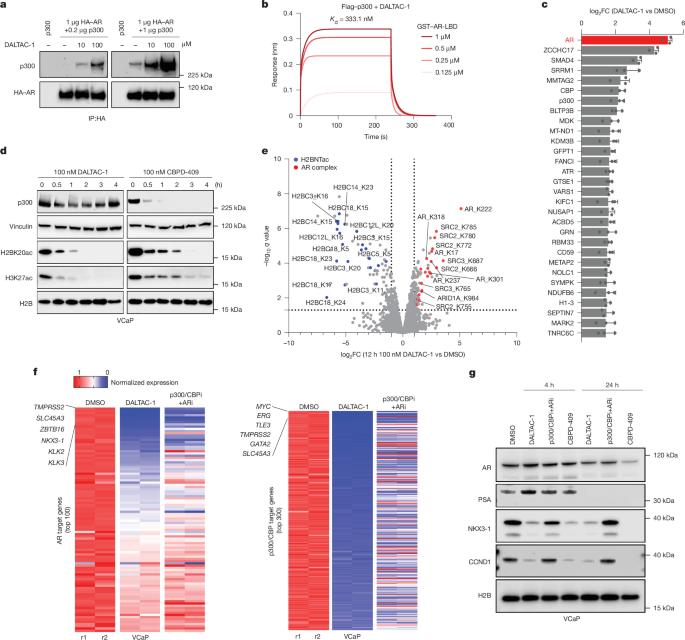
a, Immunoblot analysis showing that DALTAC-1 induced a dose-dependent interaction between purified full-length AR and p300 proteins. Purified p300 (0.2 or 1 μg) was incubated with or without purified HA–AR (1 μg) and the indicated concentrations of DALTAC-1, followed by immunoprecipitation (IP) with anti-HA (three independent experiments). b, Representative fitted BLI sensorgrams showing concentration-dependent binding of titrated GST–AR-LBD to immobilized Flag–p300 in the presence of 10 μM DALTAC-1, with an apparent Kd of 333.1 nM (three independent experiments). c, The top 30 proteins whose interactions with p300 were significantly (P < 0.05) increased in DALTAC-1-treated VCaP cells, as determined by immunoprecipitation–mass spectrometry analysis. AR is shown in red. n = 3 independently treated and processed VCaP cell samples per treatment condition. Two-sided moderated t-test with Benjamini–Hochberg correction. d, Immunoblot analysis of indicated proteins and histone marks in VCaP cells treated with 100 nM of indicated compounds for noted durations (three independent experiments). Vinculin is used as same-gel p300 loading control; H2B is a separate-gel control from the same samples. e, Volcano plot showing altered lysine acetylation levels in VCaP cells treated with 100 nM DALTAC-1 versus DMSO for 12 h. H2BNTac sites are highlighted in blue and components of the AR complex are highlighted in red. Data are plotted from n = 3 independently treated and processed samples. Statistical significance was assessed using two-sided unpaired t-tests followed by Benjamini–Hochberg correction for multiple comparisons. FC, fold change. f, RNA-seq heat maps for AR and p300/CBP target genes in VCaP cells treated with DALTAC-1 or p300/CBPi plus ARi (100 nM, 4 h). n = 2 independently treated and processed samples. g, Immunoblot analysis of indicated proteins in VCaP cells treated with 100 nM indicated compounds for 4 and 24 h. H2B is a separate-gel control from the same samples (three independent experiments).
Intracellular ternary complex formation was confirmed by ELISA, which showed that DALTAC-1, but not Neg-1 or Neg-2, enhanced AR–p300 association in VCaP cells (Extended Data Fig. 3g). Co-immunoprecipitation similarly demonstrated that DALTAC-1 enhanced the interaction between AR and p300/CBP (Extended Data Fig. 3h,i). Consistently, reciprocal immunoprecipitation–mass spectrometry analyses identified AR as strongly enriched in p300 immunoprecipitates, and p300/CBP were among the most enriched AR-associated proteins following DALTAC-1 treatment (Fig. 2c and Extended Data Fig. 3j,k). Proximity ligation assays (PLAs) further showed DALTAC-1 increased AR–p300 proximity, which was competitively blocked by either p300/CBPi or ARi (Extended Data Fig. 3l,m). Sequential co-immunoprecipitation of co-expressed Flag–AR, haemagglutinin (HA)–AR, and V5-p300 showed that DALTAC-1 maintained the interaction of HA–AR with Flag–AR from p300-containing complexes, indicating the incorporation of multiple AR molecules into the DALTAC-1-induced complex and supporting AR dimerization within the complex (Extended Data Fig. 3n). In 22Rv1 cells, DALTAC-1 enhanced p300 association with full-length AR but not the LBD-deficient AR-V7 variant, demonstrating the LBD dependence of the association (Extended Data Fig. 3o). DALTAC-1 also promoted p300 association with the DNA-binding-deficient AR-C619Y mutant35 (Extended Data Fig. 3p), indicating that complex formation does not require AR binding to DNA. Together, these findings demonstrate that DALTAC-1 induces the formation of a LBD-dependent AR–p300 complex.
Domain-resolved interaction mapping in HEK293 cells showed minimal basal association between AR and the p300 core region, comprising the BRD, RING/PHD and HAT domains, yet DALTAC-1 strongly enhanced this interaction (Extended Data Fig. 3q). Reciprocal domain mapping demonstrated that DALTAC-1 selectively recruited the AR-LBD, but not the AR-NTD, to p300 (Extended Data Fig. 3r). Accordingly, co-expressed AR-LBD and p300 core region showed minimal basal association but robust complex formation following DALTAC-1 treatment (Extended Data Fig. 3s), establishing a non-native AR-LBD–p300 core interaction.
To investigate its structural basis, we modelled the AR-LBD–DALTAC-1–p300-BRD ternary complex and the corresponding binary complexes, followed by 1-µs molecular dynamics simulations. The top-scoring ternary model predicted an AR–p300 interface stabilized by polar and hydrophobic interactions (Extended Data Fig. 3t). Key contacts included salt bridges between Asp1088 of p300 and Arg871 of AR and between Arg1133 of p300 and Glu872 of AR; hydrogen bonds involving AR Arg871–p300 Gly1085 and AR Ile816–p300 Leu1083; hydrophobic packing between Ile816 of AR and Leu1083 of p300; and a cation–π interaction between His874 of AR and the piperazine moiety of DALTAC-1. Molecular mechanics-generalized born surface area (MM-GBSA) analysis yielded a more favourable model-dependent interaction-energy estimate for the ternary complex (−161.8 ± 0.6 kcal mol−1) than for the DALTAC-1–AR (−81.4 ± 0.6 kcal mol−1) or DALTAC-1–p300 (−90.3 ± 0.4 kcal mol−1) binary complexes (Extended Data Fig. 3u,v). Computational removal of DALTAC−1 yielded an AR–p300 interface energy of −53.5 ± 1.1 kcal mol−1, consistent with favourable protein complementarity (Extended Data Fig. 3v).
Experimentally, mutation of the predicted interface residues in AR(R871A/E872A) and p300(D1088A/R1133A) substantially impaired DALTAC-1-induced complex formation (Extended Data Fig. 3w). Together, these findings establish that DALTAC-1 rewires AR–p300 domain interactions to form a stable ternary complex, providing a mechanistic basis for its downstream epigenetic and transcriptional effects.
DALTAC-1 reprograms the AR+ prostate cancer acetylome
In contrast to the p300/CBP degrader CBPD-409, DALTAC-1 did not reduce p300/CBP protein abundance but rapidly suppressed H3K27ac and H2BK20ac, two histone marks associated with active enhancers (Fig. 2d and Extended Data Fig. 4a–c). H2BNTac is deposited exclusively by p300/CBP and marks active enhancers in prostate cancer8. Our data demonstrate that DALTAC-1 exerts a de novo heterobifunctional effect by potently suppressing this critical histone mark without degrading p300/CBP. This level of H2BNTac suppression was not observed with the inactive analogues Neg-1 and Neg-2 or with the combination of AR and p300/CBP inhibitors (Extended Data Fig. 4a–c). DALTAC-1 also showed limited activity in AR-negative Kelly cells, supporting its AR-dependent selectivity (Extended Data Fig. 4d). Furthermore, in HEK293 cells, DALTAC-1-driven loss of H2BNTac increased in direct proportion to the amount of ectopically expressed AR, underscoring that both AR and p300 are required for DALTAC activity (Extended Data Fig. 4e). Moreover, a 2-h DALTAC-1 pulse caused sustained suppression of histone acetylation and MYC expression after washout, whereas the effects of combined AR and p300/CBP inhibitors were markedly reduced (Extended Data Fig. 4f).
Beyond histone substrates, p300/CBP acetylate non-histone regulators of transcription and signal transduction36. Whereas p300/CBP HAT inhibitors and degraders broadly suppress histone and non-histone acetylation7,8, acetyl-lysine profiling in VCaP cells revealed a distinct acetylation signature induced by DALTAC-1. DALTAC-1 strongly suppressed global histone acetylation, particularly enhancer-associated H2BNTac, more potently than combined p300/CBP and AR inhibition (Fig. 2e and Extended Data Fig. 4g–i). Consistently, immunoprecipitation–mass spectrometry identified H2B as the most significantly displaced p300 interactor, indicating disruption of p300 engagement with its canonical histone substrate (Extended Data Fig. 3j).
Concurrently, DALTAC-1, but not combined p300/CBP and AR inhibition, increased acetylation of AR and its cofactors SRC2 and SRC337 (Fig. 2e and Extended Data Fig. 4g,h,j,k). AR acetylation occurred in the NTD, suggesting that DALTAC-1-induced ternary complex formation redirects p300 catalytic activity towards the N terminus of AR as well as its associated cofactors. In an ARE reporter assay, glutamine substitution of the five SRC2 lysines most strongly acetylated by DALTAC-1 (multi-K-Q) modestly increased AR activity relative to wild-type SRC2, whereas alanine substitution (multi-K-A) modestly reduced it, indicating that acetylation at these sites has limited effects on SRC2 coactivator function (Extended Data Fig. 4l,m). Thus, DALTAC-1 suppresses enhancer-linked histone acetylation while selectively increasing acetylation of AR and its associated cofactors.
DALTAC-1 blocks oncogenic transcription
We next examined how DALTAC-1-mediated reprogramming of p300 substrate engagement affects transcription. DALTAC-1 strongly suppressed AR activity in an ARE–GFP reporter assay and repressed canonical AR target genes, whereas Neg-1, Neg-2 or combined p300/CBP and AR inhibition exhibited weaker effects (Extended Data Fig. 5a–c). RNA sequencing (RNA-seq) and gene set enrichment analysis (GSEA) in VCaP cells revealed downregulation of androgen response, p300/CBP target, MYC target and MTORC1 signalling pathways (Fig. 2f and Extended Data Fig. 5d–f). Chromatin immunoprecipitation with sequencing (ChIP–seq) further showed rapid RNA polymerase II (Pol II) displacement from AR and p300 target loci (Extended Data Fig. 5g). Together, unlike the transcriptional activation mechanism of TCIPs19,20, DALTAC-1 represses AR–p300/CBP-driven oncogenic transcription.
Consistently, DALTAC-1 and the p300/CBP degrader CBPD-409 markedly reduced prostate-specific antigen (PSA), CCND1 and NKX3-1 protein levels, whereas combined p300/CBP and AR inhibition had modest effects on these proteins (Fig. 2g). Quantitative proteomics confirmed selective depletion of AR- and MYC-regulated proteins without reducing AR or p300/CBP abundance (Extended Data Fig. 5h). Together, these results show that DALTAC-1 reprograms p300/CBP substrate utilization to broadly suppress AR-driven oncogenic transcription.
We next tested whether this activity requires an antagonist-derived AR binder. We replaced the AR moiety of DALTAC-1 with enobosarm38, a non-steroidal AR agonist, to generate JZY4055 (Extended Data Fig. 5i). JZY4055 potently inhibited growth of AR+ VCaP cells (Extended Data Fig. 5j). Whereas enobosarm activated canonical AR target genes, JZY4055 repressed these genes and reduced p300/CBP-dependent H2BK20ac and H3K27ac, as well as the expression of AR- and p300/CBP-regulated proteins NKX3-1 and MYC (Extended Data Fig. 5k,l). These data demonstrate that AR–p300/CBP DALTAC-mediated transcriptional repression does not require an antagonist-derived AR binder.
DALTAC-1 reshapes the AR–p300 enhanceosome
p300/CBP are central AR coactivators that sustain oncogenic transcription through enhancer-associated histone acetylation8. To investigate how DALTAC-1 modulates p300 chromatin occupancy, we performed p300 ChIP–seq in VCaP cells. DALTAC-1 markedly expanded and redistributed p300 binding genome-wide (Fig. 3a). Approximately 40% of DALTAC-1-depleted peaks were promoter-proximal, whereas gained peaks occurred predominantly in intronic and distal intergenic regions, with only approximately 10% being located near promoters, indicating increased enhancer engagement (Fig. 3b). At AR-binding enhancer sites identified under DMSO (control) conditions, DALTAC-1 proportionally increased AR and p300/CBP occupancy (Fig. 3c and Extended Data Fig. 6a,b). Motif analysis revealed that gained p300 and CBP peaks were strongly enriched for canonical palindromic AREs and closely related nuclear receptor motifs, including progesterone- and glucocorticoid-response elements (PRE and GRE)39 (Fig. 3d and Extended Data Fig. 6c,d). These findings demonstrate that DALTAC-1 redistributes p300/CBP through AR-dependent recruitment and stabilizes neomorphic AR–p300/CBP complexes on enhancer chromatin.
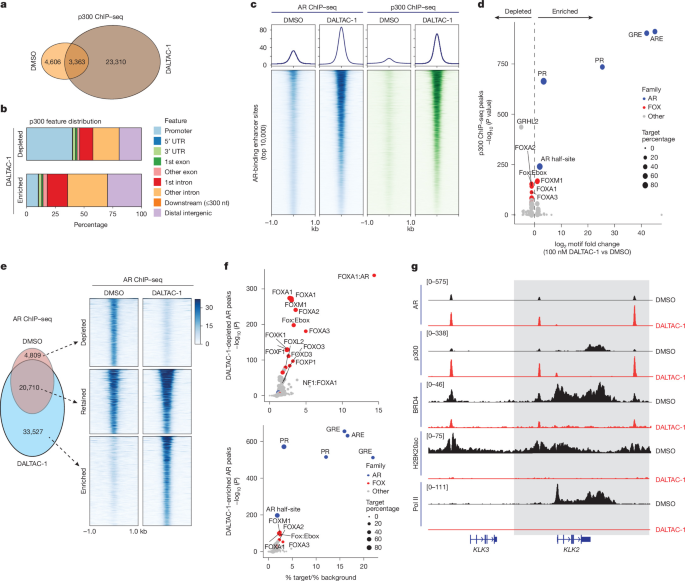
a, Venn diagram of genome-wide p300 ChIP–seq peaks in VCaP cells treated with DMSO or 100 nM DALTAC-1 for 3 h. b, Genomic distribution of p300 ChIP–seq peaks depleted or enriched by DALTAC-1, in VCaP cells treated with DMSO or 100 nM DALTAC-1 for 3 h. UTR, untranslated region. c, ChIP–seq read-density heat maps of AR and p300 at AR-bound enhancer sites in VCaP cells treated with DMSO or 100 nM DALTAC-1 for 3 h (n = 1). d, HOMER analysis showing the fold enrichment and statistical significance of sequence motifs at non-promoter p300-binding sites in VCaP cells treated with DMSO or 100 nM DALTAC-1 for 3 h. P values were calculated using the one-sided cumulative binomial test implemented in HOMER. e, Left, overlap of genome-wide AR ChIP–seq peaks in VCaP cells treated with DMSO or 100 nM DALTAC-1 for 3 h. Right, read-density heat maps of AR at DALTAC-1-depleted, retained and enriched AR-binding sites (n = 1). f, HOMER analysis of sequence motifs that are enriched at non-promoter AR-binding sites showing decreased (top) or increased (bottom) AR occupancy following treatment of VCaP cells with 100 nM DALTAC-1 for 3 h. Motif frequencies in the target and background regions and the corresponding P values are shown. P values were calculated using the one-sided cumulative binomial test implemented in HOMER. g, ChIP–seq tracks of the indicated proteins and histone marks at the KLK3–KLK2 locus in VCaP cells treated with DMSO or 100 nM DALTAC-1 for 3 h.
DALTAC-1 also significantly reshaped the AR cistrome (Fig. 3e). DALTAC-1 treatment increased AR occupancy at palindromic AREs while reducing binding at FOXA1–AR chimeric motifs associated with oncogenic AR neo-enhanceosomes40 (Fig. 3e,f and Extended Data Fig. 6e–g). Thus, rather than uniformly increasing AR binding, DALTAC-1 redirected AR away from oncogenic enhancers and concentrated it at canonical nuclear receptor response elements. Because AREs share substantial sequence similarity with PREs39, we examined whether their enrichment reflected progesterone receptor (PR) activation. DALTAC-1 instead reduced PR occupancy genome-wide while increasing AR occupancy at PR-binding sites (Extended Data Fig. 6h,i), indicating that these shared motifs were predominantly engaged by AR. Despite increased AR and p300/CBP binding, DALTAC-1 markedly decreased coactivator (BRD4 and ERG) recruitment, Pol II loading, and histone acetylation (H3K27ac and H2BK20ac) at AR/p300 co-bound enhancers (Extended Data Fig. 6j,k). DALTAC-1 therefore stabilized AR–p300/CBP chromatin binding while functionally decommissioning the associated transcriptional complexes.
Oncogenic transcription factors such as ERG and FOXA1 mutants are known to redirect AR to non-canonical AREs, thereby forming cancer-specific AR neo-enhanceosomes that drive lineage-dependent oncogene programs8,41. In TMPRSS2–ERG fusion-positive VCaP cells42, DALTAC-1 globally reduced ERG occupancy, although a subset of new ERG peaks emerged in intronic and distal intergenic regions (Extended Data Fig. 7a–d). Motif analysis revealed that DALTAC-1 depleted ERG binding at canonical ETS motifs and redistributed ERG towards palindromic AREs (Extended Data Fig. 7e). At non-promoter AR–ERG co-bound regions, DALTAC-1 increased AR and p300 occupancy, modestly increased SRC2 binding, and minimally altered FOXA1 occupancy (Extended Data Fig. 7f–h). Nonetheless, ERG and BRD4 binding declined, whereas Pol II occupancy and enhancer-associated histone acetylation were almost completely lost (Extended Data Fig. 7i). Thus, DALTAC-1 collapses AR–ERG neo-enhancers into a transcriptionally inactive state despite increased AR–p300 occupancy, dismantling the oncogenic output of the AR–ERG neo-enhanceosome (Fig. 3g and Extended Data Fig. 7j).
DALTAC-1 suppresses prostate cancer organoid growth
To assess DALTAC-1 efficacy across prostate cancer subtypes, we tested organoids derived from genetically engineered mouse models. We treated organoids derived from wild-type, Rosa26ERG/Pten−/−, Pten−/−, FOXA1 R265–71del+/+/Trp53−/− and Trp53−/− mouse prostates with DALTAC-141,43. DALTAC-1 preferentially reduced the number and size of Rosa26ERG/Pten−/− and FOXA1 R265–71del+/+/Trp53−/− organoids and suppressed MYC and CCND1 relative to wild-type, Pten−/− and Trp53−/− organoids (Extended Data Fig. 8a–d). Given that ERG and mutant FOXA1 enhance AR activity and sustain the AR neo-enhanceosome5,41,43,44, these findings suggest that DALTAC-1 effectively suppresses AR-driven prostate cancer. Furthermore, we evaluated DALTAC-1 efficacy across organoids derived from patient-derived xenograft (PDX) tumours45,46. Consistently, DALTAC-1 potently inhibited AR+ organoids, particularly AR+/ERG+ models (Fig. 4a). Thus, DALTAC-1 exhibits potent, lineage-selective activity across mouse and human organoid models.
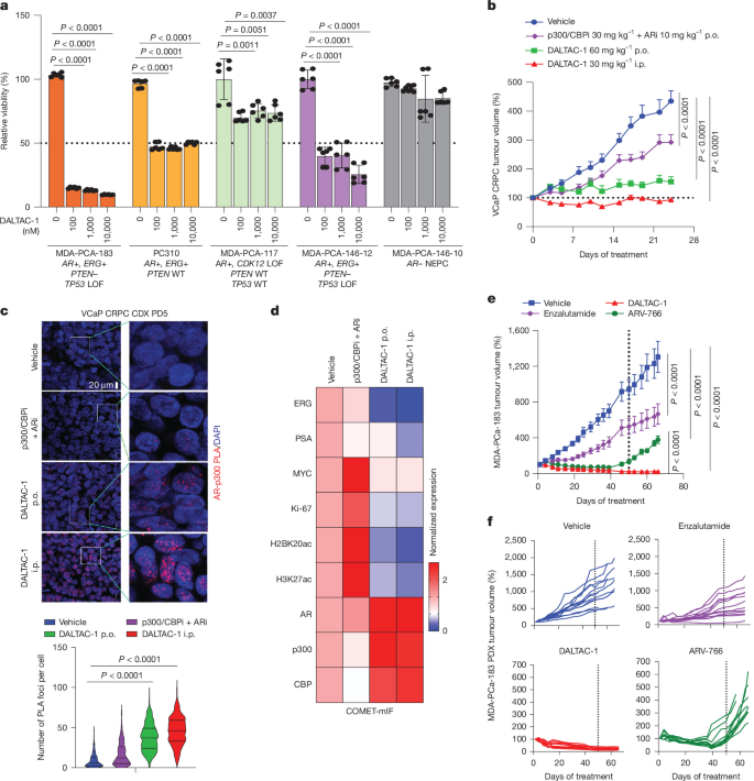
a, Viability of PDX-derived human organoids treated with DMSO or DALTAC-1 (100, 1,000 or 10,000 nM) for 14 days. Data are mean ± s.d. from three independent organoid culture wells per condition (n = 3 biological replicates), each divided into two wells for 3D-CTG measurement (2 technical replicates; 6 measurements shown). P values were calculated using two-sided t-tests. LOF, loss of function; NEPC, neuroendocrine prostate cancer; WT, wild type. b, VCaP-CRPC tumour volumes after vehicle, DALTAC-1 (30 mg kg−1 intraperitoneal administration (i.p.) or 60 mg kg−1 p.o. (oral administration); 5 times weekly in week 1, then 3 times weekly for 3 weeks), or p300/CBPi plus ARi (30 mg kg−1 p300/CBPi plus 10 mg kg−1 ARi orally, 5 times weekly). Data are mean ± s.e.m.; n = 14 tumours for vehicle, oral DALTAC-1 and p300/CBPi plus ARi, and n = 16 tumours for intraperitoneal DALTAC-1. End-point volumes were analysed by two-sided t-test. c, Top, representative images of AR–p300 PLA assays in PD5 tumour samples from the VCaP-CRPC study. Bottom, quantification of AR–p300 PLA foci per cell (n = 155 cells for vehicle, n = 204 cells for ARi plus p300/CBPi, n = 187 cells for DALTAC-1 orally, n = 174 cells for intraperitoneal DALTAC-1). P values were calculated using a two-sided t-test. In violin plots, the central line is the median; dotted lines represent 25th–75th percentiles; violins represent kernel density estimates; whiskers extend to minimum and maximum values. CDX, cell line-derived xenograft. d, Heat map showing normalized median expression levels of the indicated targets from PD5 tumours in the VCaP-CRPC study using COMET-mIF. e, MDA-PCa-183 PDX growth during treatment with vehicle, DALTAC-1 (30 mg kg−1 intraperitoneally, 3 times weekly), ARV-766 (10 mg kg−1 orally, 5 times weekly) or enzalutamide (10 mg kg−1 orally, 5 times weekly). Dosing stopped on day 50, followed by 16 days of monitoring (mean ± s.e.m.; n = 12 tumours for vehicle, DALTAC-1 and enzalutamide, and n = 11 tumours for ARV-766; two-sided t-test). f, Graph depicting the tumour volume curves of individual tumours from e.
Potent in vivo efficacy of DALTAC-1 in prostate cancer
To evaluate in vivo activity of DALTAC-1, we first examined VCaP castration-resistant prostate cancer (VCaP-CRPC) xenografts established in castrated male severe combined immunodeficiency (SCID) mice (Extended Data Fig. 9a). Mice were randomized to DALTAC-1 administered intraperitoneally (30 mg kg−1) or orally (60 mg kg−1), or an equipotent combination of p300/CBPi and ARi. Intraperitoneal DALTAC-1 induced tumour regression in 75% (12 out of 16) of tumours, whereas oral dosing also robustly suppressed tumour growth and outperformed the combined inhibitors (Fig. 4b and Extended Data Fig. 9b–d). In vivo PLA confirmed increased AR–p300 proximity in treated tumours (Fig. 4c). COMET multiplex immunofluorescence (COMET-mIF) analyses revealed that DALTAC-1 reduced ERG, PSA, MYC, Ki-67, H3K27ac and H2BK20ac without decreasing AR, p300 or CBP abundance, consistent with non-degradative AR–p300/CBP rewiring (Fig. 4d and Extended Data Fig. 9e,f).
We next assessed the efficacy of DALTAC-1 in two prostate cancer PDX models, MDA-PCa-146-12 and MDA-PCa-18345,47. DALTAC-1 significantly inhibited MDA-PCa-146-12 growth (Extended Data Fig. 9g) and induced rapid, sustained regression of TMPRSS2–ERG-positive, PTEN-deficient MDA-PCa-183 tumours, with no progression observed during the 35-day treatment period (Extended Data Fig. 9h–k). In the MDA-PCa-183 model, DALTAC-1 induced marked tumour regression throughout the 50-day treatment period, whereas enzalutamide produced only modest tumour growth inhibition and ARV-766 was less effective than DALTAC-1 (Fig. 4e,f). Following treatment withdrawal, DALTAC-1-treated tumours showed no evident recurrence for 16 days, whereas ARV-766-treated tumours regrew (Fig. 4e,f). DALTAC-1 was well tolerated, with no significant changes in body weight, liver or kidney function, or haematological parameters (Extended Data Fig. 10a–e). Together, DALTAC-1 produces potent and durable antitumour responses across prostate cancer models through selective, non-degradative reprogramming of AR–p300/CBP complexes.
Discussion
Recent advances in CIP have led to the development of a diverse array of compounds capable of modulating protein functions through enforced protein–protein interactions14. RIPTACs induce proximity between a target protein and a pan-essential effector that disrupts the function of the essential effector, leading to selective cancer cell death while sparing healthy cells17. By contrast, TCIPs recruit transcriptional coactivators to specific genomic loci, paradoxically activating the target pathway by enhancing transcription of pro-apoptotic genes that are normally repressed by corepressors such as BCL619,20. The emerging clinical activity of daraxonrasib (RMC-6236), which forms a ternary complex with cyclophilin A and active RAS, further supports the translational potential of proximity-induced pharmacology48. Recent molecular glue degraders further demonstrate that small molecules can stabilize pre-existing or newly enabled protein interfaces and alter target organization to promote E3 ligase-dependent degradation49. Complementary studies targeting FOXA1, SWI/SNF and p300 establish that DNA-binding specificity, ATPase cycling and chromatin engagement are also pharmacologically actionable50,51,52. DALTACs build on these principles but produce a distinct pharmacological outcome: rather than recruiting an E3 ligase, degrading a complex component or modulating an individual regulator, a DALTAC bifunctional molecule imposes an additional domain-selective interaction between two members within an existing transcriptional complex, converting the complex into a non-productive state. Our proof-of-concept compound, AR–p300/CBP DALTAC-1, targets the AR–p300/CBP complex by repurposing the natural interfaces to block activation of the AR oncogenic enhanceosome. Mechanistically, AR–p300/CBP DALTAC-1 potently suppresses AR transactivation by disrupting the chromatin landscape at AR-bound enhancer sites, particularly AR/ERG co-bound neo-enhancer sites, as evidenced by reduced cofactor recruitment, loss of histone acetylation marks, and displacement of Pol II. Functionally, this shutdown of AR neo-enhanceosome activity translates into strong antitumour efficacy in AR+ prostate cancer models, underscoring the translational potential of DALTAC-1 as a precision therapeutic agent.
The activity of CIP molecules is founded on formation of a ternary complex between the recruited proteins. Using purified full-length proteins, we showed that DALTAC-1 directly enhances AR–p300 complex formation in the absence of DNA or accessory factors. This association requires simultaneous engagement of both proteins by the intact bifunctional molecule and cannot be reproduced by unlinked ligands or control compounds lacking either AR- or p300-binding activity. The activities of multi-protein transcriptional complexes are governed by finely tuned protein–protein interactions at the sub-domain level53. AR normally recruits transcriptional coactivators primarily through its intrinsically disordered NTD, whereas transcription factors engage distinct regions of p300, including its N- and C-terminal regions, to support histone acetylation and transcriptional activation2,9. Our domain-mapping and interface-mutagenesis studies support a refined model in which DALTAC-1 does not necessarily abolish these native contacts. Rather, it imposes an additional, neomorphic interaction between the AR-LBD and the p300 core region containing the BRD and HAT domain, thereby reorganizing the domain-level engagement within the retained AR–p300 complex. Although the atomic architecture of the ternary complex remains unresolved, owing in part to the large size, conformational flexibility and extensive intrinsically disordered regions of full-length AR and p300, our orthogonal biochemical and cellular data demonstrate a DALTAC-induced binding pattern that is distinct from the basal complex and functionally alters AR–p300/CBP activity. This interaction reprogramming markedly suppresses histone acetylation, including H2BNTac, despite the inability of the p300-binding moiety alone to directly inhibit this modification8. In parallel, DALTAC-1 induces acetylation of the AR-NTD and core components of the AR complex, including SRC2/3 and ARID1A/B4,37,54. Because p300 has previously been shown to acetylate AR primarily within the hinge region under androgen stimulation55, the emergence of AR-NTD acetylation suggests that DALTAC-1 redirects p300 substrate engagement, potentially by bringing the AR-NTD into altered proximity to the p300 HAT domain. This substrate reprogramming distinguishes DALTAC-1 from conventional p300/CBP inhibitors and degraders: unlike BRD inhibitors, it extinguishes enhancer-associated H2BNT acetylation and broadly represses transcription, and unlike PROTAC degraders, it acts independently of ubiquitin–proteasome-mediated target depletion.
Because many oncogenes also have essential roles in normal physiology, their pan-inhibition often causes severe side effects. For example, systemic inhibition of p300/CBP disrupts haematopoiesis and leads to thrombocytopenia13. Thus, achieving lineage-selective targeting of oncogenic pathways is an urgent therapeutic need. Existing approaches such as antibody–drug conjugates deliver cytotoxic payloads selectively to cells that express target surface antigens56. However, their broader applicability is limited by the restricted repertoire of tumour-specific surface markers and, in many cases, by suboptimal pharmacokinetic properties57. By contrast, DALTACs offer a fundamentally distinct strategy, selectively rewiring lineage-specific protein complexes that drive oncogenic transcription. In this study, AR–p300/CBP DALTAC-1 specifically inhibits the growth of AR+ prostate cancer cells with potency comparable to or better than the p300/CBP degrader CBPD-409 but shows markedly reduced activity in p300/CBP-dependent, AR-negative cancers such as neuroblastoma, Ewing sarcoma and haematological malignancies8. Notably, in AR-negative cells, DALTAC-1 does not suppress H2BNTac, underscoring that DALTAC-1-mediated inhibition of p300/CBP histone acetylation is context-dependent and conferred by domain reprogramming within AR–p300/CBP complexes.
In summary, we establish DALTACs as a new type of chemical proximity modulators that rewire domain-specific protein interactions to selectively deactivate oncogenic complexes. DALTAC-1 induces formation of an AR–p300–DALTAC-1 ternary complex and reprograms the substrate engagement of p300/CBP, markedly suppressing global histone acetylation while enhancing the acetylation of core components within the AR complex. Consequently, DALTAC-1 reinforces AR–p300/CBP co-occupancy at palindromic ARE loci but disrupts the higher-order assembly of the AR complex and erases histone activation marks at AR neo-enhanceosomes (Fig. 5). This work expands the conceptual framework of induced-proximity therapies beyond degradation and coactivator hijacking, paving the way for the rational design of domain-alteration strategies for precise control of disease-relevant protein complexes.
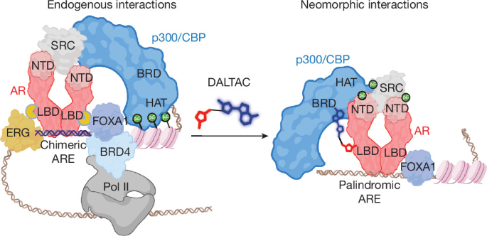
Left, endogenous AR–p300/CBP interactions recruit coactivators and promote histone acetylation at oncogenic AR-bound enhancers, sustaining oncogenic transcription. Right, DALTAC-1 induces a neomorphic interaction between AR and p300/CBP that stabilizes the ternary complex at palindromic AREs and reprograms p300/CBP substrate engagement, suppressing histone acetylation while increasing acetylation of core AR complex components. Consequently, DALTAC-1 disrupts productive complex assembly at AR neo-enhanceosomes, erases activating histone marks and suppresses oncogenic transcription. Created in BioRender; Young, E. https://BioRender.com/3ei5yd8 (2026).
Methods
Cell lines
VCaP, LNCaP, 22Rv1, DU145, PC-3, MCF-7, HepG2, A549, K562, HL-60, U937, HEK293, WPMY-1, U-87 MG, BJAB, SK-ES-1, RD-ES, A204, PBMC, NCI-H526, AU565, MM1.S, SU-DHL-8 and NCI-H211 were purchased from American Type Culture Collection (ATCC). MOLT-16, NALM6, REH and TALL-1 were obtained from the German Collection of Microorganisms and Cell Cultures (DSMZ). CHLA-99, CHLA-258, CHLA-32, CB-AGPN, COG-E-352 and TC-205 were obtained from the Children’s Oncology Group (COG) Cell Line and Xenograft Repository. GM24694 was obtained from the Coriell Institute for Medical Research. PNT2 and MFM223 were obtained from Sigma-Aldrich. The Kelly cell line was obtained from European Collection of Authenticated Cell Cultures (ECACC). CADOES1 was obtained from Cell Lines Service (CLS). LK2 was obtained from the Japanese Collection of Research Bioresources (JCRB) Cell Bank. NB-1 was obtained from Sekisui XenoTech.
CWR-R1 cells (Sigma-Aldrich, SCC118) were provided by D. Vander Griend. SU-DHL-5 cells (ATCC CRL-2958/DSMZ ACC 571) were provided by the R. Ryan laboratory. TC-32 cells (COG Cell Line and Xenograft Repository) were provided by the E. R. Lawlor laboratory. T47D cells (ATCC HTB-133) were provided by the C. Speers Laboratory. SUM185PE cells (Asterand Bioscience) were provided by the Rae laboratory. WA72 is an in-house PDX-derived cell line established at the Michigan Center for Translational Pathology (MCTP), University of Michigan, and is not commercially available. All cell lines were cultured according to conditions recommended by the originating repository or providing laboratory, as appropriate.
Two cell lines used in this study, BJAB and U937, are listed in the International Cell Line Authentication Committee (ICLAC) Register of Misidentified Cell Lines. Both entries reflect historical contamination reports that have since been resolved. U-87 MG (obtained from ATCC) is also listed in the ICLAC Register (ICLAC-00535). This cell line was included at the specific request of a peer reviewer, to test compound activity in an AR+, non-prostate cancer cellular context, given reports that AR functions as an oncogenic driver in this model. This experiment was intended to provide broader context on compound selectivity across AR+ tumour types outside of prostate cancer, and not to draw conclusions regarding glioblastoma tissue-of-origin biology specifically. No other cell lines used in this study appear in the ICLAC Register.
All human cell lines were authenticated by short tandem repeat (STR) genetic testing. All cell lines were tested biweekly to be free of mycoplasma contamination.
Chemistry
Detailed synthesis and chemical data for all designed DALTAC molecules and negative controls are provided in the Supplementary Methods.
Antibodies
For immunoblotting, the following antibodies were used: p300 (Invitrogen: MA1-16608, 1:500); AR (Abcam: ab133273, 1:1,000); H2BK20ac (Cell Signaling Technology: 34156S, 1:1,000); H2BK16ac (Abcam: ab177427, 1:1,000); H3K27ac (Cell Signaling Technology: 8173S, 1:1,000); H2B (Active Motif: 39210, 1:1,000); H3 (Cell Signaling Technology: 3638S, 1:1,000); MYC (Cell Signaling Technology: 9402S, 1:1,000); KLK3/PSA (Dako: A0562, 1:1,000); NKX3-1 (Cell Signaling Technology: 83700S, 1:1,000); CCND1 (Abcam: ab16663, 1:1,000); vinculin (Cell Signaling Technology: 18799S, 1:1,000); GAPDH (Santa Cruz Biotechnology: sc-47724, 1:1,000); BRD4 (Bethyl Laboratories: A700-004CF, 1:1,000); AR-V7 (Cell Signaling Technology: 19672S, 1:1,000); HA tag (Cell Signaling Technology: 3724S, 1:1,000); CBP (Abcam: ab253202, 1:1,000); and V5 (Invitrogen, R960-25, 1:1,000). Horseradish peroxidase (HRP)-conjugated goat anti-rabbit (Bio-Rad; 1:10,000) and goat anti-mouse (Bio-Rad; 1:5,000) secondary antibodies were used.
For ChIP–seq, the following antibodies were used: H3K27ac (Diagenode: C15410196, 1 µg per million cells); p300 (Active Motif: 61401, 2 µg per million cells); H2BK20ac (Cell Signaling Technology: 34156S, 1 µg per million cells); AR (Abcam: ab108341, 2 µg per million cells); FOXA1 (Thermo Fisher Scientific: PA5-27157, 2 µg per million cells); BRD4 (Diagenode: C15410337, 2 µg per million cells); ERG (Cell Signaling Technology: 97249S, 2 µg per million cells); RNA Pol II (Diagenode: C15200253, 1 µg per million cells); CBP (Abcam: ab253202, 2 µg per million cells); SRC2 (Cell Signaling Technology: 96687S, 4 µl per million cells) and PR (Cell Signaling Technology: 8757S, 4 µl per million cells).
For immunoprecipitation, acetylated-lysine antibody (Cell Signaling Technology: 9441S) was used at 1 µg per 100 µg of lysate.
For immunofluorescent staining (COMET-mIF) and PLA assay, the following antibodies were used: p300 (Invitrogen: 33-7600, 1:150); CBP (Invitrogen: PA5-27369, 1:150); AR (Abcam: ab133273, 1:400); H3K27ac (Cell Signaling Technology: 8173S, 1:100); Ki-67 (Abcam: ab243878, 1:300); H2BK20ac (Abcam: ab177430, 1:250); CK8 (Abcam: ab53280, 1:50); ERG (Abcam: ab92513, 1:200); PSA (Cell Marque: 324M-16, 1:50); and MYC (Abcam: ab32072, 1:50).
For ELISA, the following antibodies were used: p300 (Cell Signaling Technology: 86377S, 1:1,000); and AR (Thermo Fisher Scientific: MA5-13426, 1:50).
Cell viability assay
VCaP cells were seeded at a density of 10,000 cells per well, and all other prostate cancer cell lines were seeded at 3,000 cells per well in poly-d-lysine-coated 96-well plates (Corning) and maintained at 37 °C with 5% CO2. After 24 h, cells were treated with a serial dilution of test compounds, with 6 replicate wells for each concentration. Following 120 h of incubation, cell viability was determined using the CellTiter-Glo Luminescent Cell Viability Assay (Promega) according to the manufacturer’s instructions. Luminescence was measured on an Infinite M1000 Pro plate reader (Tecan) and data were analysed using GraphPad Prism (v10; GraphPad Software).
Incucyte proliferation assays
A total of 4,000 VCaP cells or 1,500 LNCaP cells per well were seeded into poly-d-lysine-coated 96-well plates. After 24 h of incubation, cells were treated with a fixed concentration (100 nM) or serial dilutions of compounds and incubated for 4 h. The medium was then removed, and cells were washed three times with phosphate-buffered saline (PBS) before fresh medium containing either vehicle or compounds was added. Phase-contrast images were collected every 4 h, and cell proliferation was quantified by measuring the percentage of confluence (phase object area).
Immunoblotting
Cells were lysed in RIPA buffer (Thermo Fisher Scientific) supplemented with 1× Halt Protease Inhibitor Cocktail (Thermo Fisher Scientific) and denatured in NuPAGE 1× LDS sample buffer containing a reducing agent (Invitrogen) by incubation at 70 °C for 15 min. Protein concentrations were determined using the Pierce BCA Protein Assay Kit (Thermo Fisher Scientific). Equal amounts of protein (15–30 µg) were resolved on NuPAGE 3–8% Tris-Acetate or 4–12% Bis-Tris protein gels (Thermo Fisher Scientific) and transferred onto 0.45-µm nitrocellulose membranes (Thermo Fisher Scientific) using the iBlot 3 Dry Blotting System (Thermo Fisher Scientific). Membranes were blocked in TBS-T (Tris-buffered saline with 0.1% Tween-20) containing 5% non-fat dry milk for 1 h at room temperature and incubated overnight at 4 °C with primary antibodies. After washing, membranes were incubated with HRP-conjugated secondary antibodies and imaged using an Odyssey CLx Imager (LI-COR Biosciences). Supplementary Figure 1 shows the full, uncropped blots with molecular weight markers for all immunoblot experiments.
Fluorescence polarization binding assays
The binding affinities of test compounds to AR and CBP/p300 were measured using fluorescence polarization-based competitive binding assays. AR binding was assessed using the PolarScreen Androgen Receptor Competitor Assay Kit (Thermo Fisher Scientific, A15880) following the manufacturer’s instructions.
CBP/p300 fluorescence polarization assays were performed in 96-well plates (MTP96VPWB-EP, Eppendorf) using a CLARIOstar microplate reader (BMG Labtech). Serial dilutions of test compounds were incubated with fluorescein-labelled tracer (2 nM final) and CBP/p300 protein (20 nM final) in assay buffer (PBS pH 7.4 supplemented with 0.01% BGG, 0.01% Tween-20, and 2 mM DTT) to a final reaction volume of 100 µl per well. Plates were mixed for 15 min and incubated at room temperature for 1 h to reach equilibrium. Fluorescence polarization (mP units) was measured at 485 nm excitation and 530 nm emission.
For both AR and CBP/p300 assays, data analysis was performed in GraphPad Prism 10 using nonlinear regression. IC50 values were determined by fitting competition curves, and inhibition constant (Ki) values for competitive inhibitors were derived using nonlinear regression based on the Kd of the tracer and the concentrations of tracer and protein in the reaction. All fluorescence polarization experiments were conducted in duplicates across three independent biological replicates.
Cellular thermal shift assay
VCaP or LNCaP cells were seeded in poly-d-lysine-coated 100 mm culture dishes (Corning) and incubated overnight. Cells were incubated with the DMSO control or indicated compounds for 4 h at 37 °C before being collected and washed once with PBS. The cell suspension was then distributed into PCR tubes (50 µl per tube) and exposed to a predefined temperature gradient for 3 min using a Veriti Thermal Cycler (Applied Biosystems). Following equilibration to room temperature, cells were lysed by three freeze–thaw cycles in liquid nitrogen. Insoluble protein aggregates were pelleted by centrifugation at 20,000g for 20 min at 4 °C, and the resulting supernatants containing soluble proteins were recovered for western blot analysis.
Computational modelling
All modelling and simulations were performed with Schrödinger 2025-2 (Schrödinger). Because co-crystal structures of the LBD of AR in an open antagonist conformation were not available, we used our previously reported pseudo-open model58. The bound AR ligand was modified to generate JZY3221, the AR ligand used in our design of DALTACs. To model the p300-BRD interaction with GNE-049, we used the CBP-BRD–GNE-781 co-crystal structure (Protein Data Bank (PDB) 5W0E) as the starting structure59. CBP and p300 are highly homologous proteins, especially in their BRDs, which share conserved sequence and structures. Both proteins were prepared for further modelling using the Protein Preparation Wizard60.
We used the following structures as inputs to generate the initial AR–DALTAC-1–p300 complex: (1) AR in complex with JZY3221; (2) the p300 complex with GNE-049; and (3) JZY3032 (DALTAC-1) prepared with LigPrep module61. These inputs were submitted to Generate Degrader Complex (default settings: MCMM 10,000 steps, maximum clashes 5, maximum poses 50) to construct the ternary AR–JZY3032–p300 complex. The top 20 predicted complexes were further refined using Prime62. The top-scoring ternary complex was then selected for molecular dynamics simulations.
To perform molecular dynamics simulations, the predicted ternary complex was solvated in an explicit water box with periodic boundary conditions. A cubic box was constructed with a 10 Å buffer between solute and box edge and filled with TIP4P water63. The OPLS4 force field was used for all components. Production dynamics used a RESPA integrator with a 2 fs time step. Temperature was maintained at 300 K with a Nosé–Hoover chain thermostat64 (relaxation time 1.0 ps). Pressure was controlled at 1 atm with a Martyna–Tobias–Klein barostat65 (relaxation time 2.0 ps). Long-range electrostatics used a 9 Å real-space cutoff with particle–mesh Ewald for reciprocal space66. One continuous 1-µs simulation was performed without restraints.
Root-mean-square deviation analysis of protein backbones and ligand position indicated that the system equilibrated after approximately 750 ns. Accordingly, trajectory analysis focused on the last 250 ns. Protein–protein interface features were defined as minimum heavy-atom distances between AR and p300. Contacts shorter than 0.5 nm (5 Å) were retained. Principal component analysis was applied to these interface distances, and components explaining 95% of the variance were kept. The reduced data were clustered with HDBSCAN (min_cluster_size 25, centroids enabled)67. For each cluster, the frame closest to the centroid in principal component analysis space was saved as a representative PDB. Cluster occupancies are reported as percentages of frames per cluster. Protein–protein interaction fingerprints were computed with Schrödinger’s analyze_trajectory_ppi.py. Clustering in this reduced dimensional space revealed two dominant populations, with the most prevalent state comprising nearly 80% of the ensemble. The interaction analysis was performed on snapshots representative of this dominant state.
MM-GBSA calculations
MM-GBSA binding energies were estimated using Prime MM-GBSA (Schrödinger Release 2025-2; thermal_mmgbsa protocol) with the OPLS4 force field and the VSGB 2.0 implicit-solvent model68,69,70. Snapshots were sampled evenly from the final 250 ns of each trajectory, after which water molecules and ions were removed and each complex, receptor, and ligand were locally energy-minimized. The MM-GBSA binding energy was calculated as ΔGbind = Ecomplex − Ereceptor − Eligand, where ΔGbind is the estimated MM-GBSA binding energy and Ecomplex, Ereceptor and Eligand are the calculated MM-GBSA energies of the complex, receptor and ligand, respectively. For compound binding, DALTAC-1 was defined as the ligand and AR, p300, or AR + p300 as the receptor. For p300 association with the ternary complex, p300 (residues 1049–1160) was defined as the ligand and AR + DALTAC-1 as the receptor. To isolate the direct AR–p300 contribution in the induced geometry, DALTAC-1 was deleted from the same ternary snapshots, with p300 defined as the ligand and AR as the receptor. MM-GBSA values were used as model-dependent interaction-energy estimates to compare the relative stability of the modelled complexes and were not interpreted as experimental binding affinities.
Full-length AR protein expression and purification
AR protein was expressed according to the manufacturer’s protocol for the Expi293 PRO Expression System Kit (Thermo Fisher Scientific, A40003643). In brief, Expi293 PRO cells were transfected with the HA–AR expression plasmid and cultured for 24 h before the addition of 1 μM R1881. The cultures were then incubated for an additional 24 h, and cells were collected 48 h post-transfection. Cell pellets were washed and collected by centrifugation at 5,000 rpm for 10 min at 4 °C. The cells were resuspended in lysis buffer containing 50 mM Tris-HCl (pH 8.0), 150 mM NaCl, 100 nM R1881 and 0.5% NP-40, supplemented with cOmplete EDTA-free Protease Inhibitor Cocktail (Roche, 4693159001). Cells were lysed by sonication, and the lysates were clarified by centrifugation at 15,000 rpm for 40 min at 4 °C. The clarified lysate was incubated with anti-HA magnetic beads (Thermo Scientific, 88837) for 3 h at 4 °C. After binding, the beads were washed three times with wash buffer containing 50 mM Tris-HCl (pH 8.0), 150 mM NaCl, and 0.05% NP-40. Bound AR protein was eluted twice using elution buffer (50 mM Tris-HCl, pH 8.0, 150 mM NaCl, 100 nM R1881 and 0.05% NP-40) supplemented with 2 mg ml−1 HA peptide (Thermo Scientific, 26184). The eluted protein was further purified by size-exclusion chromatography using a Superdex 200 Increase 10/300 GL column (GE Healthcare) pre-equilibrated with gel filtration buffer containing 50 mM Tris-HCl (pH 8.0), 150 mM NaCl, 100 nM R1881, and 0.05% NP-40. Peak fractions containing purified AR protein were pooled to a final concentration of approximately 0.5 mg ml−1 and used for all subsequent studies.
Purified-protein AR–p300 immunoprecipitation assay
For each reaction, 15 μl of anti-HA magnetic beads (Thermo Fisher Scientific, 88837) was blocked with 0.3% BSA in PBS for 1 h at 4 °C and washed three times with AR protein buffer (50 mM Tris-HCl, pH 8.0; 150 mM NaCl; 0.05% NP-40) for 5 min per wash. Purified HA-tagged full-length AR (1 μg) was incubated with the blocked beads for at least 4 h at 4 °C. The beads were then washed three times with p300 protein buffer (25 mM HEPES-NaOH pH 7.5, 300 mM NaCl, 0.04% Triton X-100, and 0.5 mM TCEP) for 10 min per wash and incubated overnight at 4 °C with purified Flag-tagged p300 (Active motif, 81858) (200 ng or 1 μg) in 350 μl of p300 protein buffer containing the indicated compounds or an equivalent volume of DMSO. Beads were subsequently washed at least three times with p300 protein buffer containing the corresponding compounds for 10 min per wash. Bound proteins were eluted in SDS–PAGE loading buffer and analysed by immunoblotting.
Ternary complex formation by BLI
Anti-Flag IgG biosensors (Sartorius, 18-5187) were pre-equilibrated in assay buffer containing 25 mM HEPES-NaOH (pH 7.5), 300 mM NaCl, 10% glycerol, 0.04% Triton X-100, and 0.5 mM TCEP. N-terminal Flag-tagged full-length recombinant p300 protein (1 μg; Active Motif, 81858) was immobilized onto the biosensors at 25 °C until the binding signal reached a plateau. After immobilization, the biosensors were washed in assay buffer to remove unbound protein and minimize nonspecific binding.
The biosensors were then incubated with recombinant GST–His-tagged AR-LBD protein (Invitrogen, A15675) at varying concentrations in assay buffer containing either 10 μM DALTAC-1 or an equivalent volume of DMSO. The association phase was monitored, followed by transfer of the biosensors into assay buffer to measure dissociation. Binding data were analysed using Octet Analysis Studio 13.0 (Sartorius). Sensorgrams obtained in the presence of DMSO were used as reference controls and subtracted from those obtained with DALTAC-1. The reference-subtracted data were globally fitted to a 1:1 binding model to determine the equilibrium dissociation constant (Kd).
Proximity ligation assay
PLA was performed according to the manufacturer’s instructions (Navinci Diagnostics) using the Naveni TriFlex Cell kit for cultured cells (AR–p300) and the NaveniFlex Tissue Red kit for VCaP-CRPC tumour sections.
Cultured cells (Naveni TriFlex Cell) were fixed, permeabilized and blocked per kit protocol instructions. Primary antibodies against AR (1:400) and p300 (1:200) were applied in antibody diluent and incubated as recommended. After washes, Naveni proximity probes were added, followed by ligation and rolling-circle amplification steps according to the vendor’s workflow. Slides were counterstained with DAPI and mounted in antifade medium. PLA signals were visualized on a Zeiss LSM900 fluorescence microscope under identical exposure settings across conditions.
For formalin-fixed paraffin-embedded (FFPE) tumour sections (NaveniFlex Tissue Red), sections were deparaffinized, rehydrated and subjected to heat-induced epitope retrieval as specified by the kit. After blocking, primary antibodies against AR (1:1,000) and p300 (1:100) were applied in antibody diluent and incubated per protocol. Following washes, NaveniFlex proximity probes, ligation and amplification were performed according to the manufacturer’s instructions. Nuclei were counterstained with DAPI, slides were mounted and images were acquired on a Zeiss LSM900 microscope using matched acquisition parameters.
Where indicated, PLA puncta were quantified as foci per nucleus using standardized thresholds across groups.
Ternary complex ELISA assay
VCaP cells were plated in 96-well plates (50,000 cells per well) and cultured for 3 days in medium containing 5% charcoal-stripped FBS. Cells were treated with a serial dilution of DALTAC-1, Neg-1, or Neg-2 compounds for 3 h. Following treatment, cells were lysed in 1× AlphaLISA SureFire Ultra Lysis Buffer (PerkinElmer) for 30 min at 4 °C with gentle mixing.
For detection, high-binding MaxiSorp 96-well plates (Thermo Fisher Scientific) were coated overnight at 4 °C with 50 µl of anti-AR antibody (clone AR441, Thermo Fisher Scientific) diluted 1:50 in PBS. After washing with TBS-T and blocking with 5% BSA for 2 h at room temperature, 50 µl of cell lysate was added to each well and incubated for 1 h at room temperature. Plates were washed three times with TBS-T and then incubated overnight at 4 °C with rabbit anti-p300 (D8Z4E, 86377, Cell Signaling Technology) antibody diluted 1:1,000 in blocking buffer.
After washing, HRP-conjugated goat anti-rabbit secondary antibody (Bio-Rad, 1706515; 1:10,000 dilution) was added and incubated for 1 h at room temperature. Plates were washed seven or eight times with TBST, and chemifluorescent signals were developed using QuantaRed Enhanced Chemifluorescent HRP Substrate (Thermo Fisher Scientific). Fluorescence (excitation 570 nm, emission 585 nm) was immediately measured using a Tecan Infinite M1000 Pro plate reader, with readings taken at 5 and 15 min.
Nuclear immunoprecipitation
Cells (50 million) were washed once with ice-cold PBS, scraped, and pelleted at 500g for 5 min at 4 °C. The cell pellet was resuspended in 2 ml hypotonic buffer (20 mM HEPES-KOH pH 7.5, 10 mM KCl, 1 mM MgCl2, 0.1% Triton X-100, 15 mM β-mercaptoethanol) supplemented with protease and phosphatase inhibitors and rotated for 1 h at 4 °C. Nuclei were collected by centrifugation at 2,000g for 5 min at 4 °C, the supernatant (cytoplasmic fraction) was discarded, and nuclei were washed once with hypotonic buffer.
The nuclear pellet was lysed in 1 ml nuclear lysis buffer (20 mM HEPES-KOH pH 7.5, 150 mM NaCl, 10 mM KCl, 1 mM MgCl2, 0.5% Triton X-100, 15 mM β-mercaptoethanol) containing protease and phosphatase inhibitors and rotated for 1 h at 4 °C. Benzonase (500 U ml−¹; Sigma-Aldrich E1014) was added during lysis to digest nucleic acids. Lysates were clarified by centrifugation at 16,000g for 20 min at 4 °C, and the supernatant was collected and precleared with 50 µl Dynabeads Protein G magnetic beads (Thermo Fisher 10004D). Protein concentration was determined using the Pierce 660 nm Protein Assay (Thermo Fisher 22660).
For each immunoprecipitation, 400 µg of precleared nuclear lysate was incubated overnight at 4 °C with either anti-androgen receptor antibody (Abcam ab108341) or anti-p300 antibody (Active Motif 61401). Antigen–antibody complexes were captured with 30 µl Dynabeads Protein G for 2 h at 4 °C. Beads were washed three times with wash buffer (20 mM HEPES-KOH pH 7.5, 150 mM NaCl, 10 mM KCl, 1 mM MgCl2).
For immunoblotting, bound proteins were eluted in 1× NuPAGE LDS Sample Buffer (Thermo Fisher NP0007) supplemented with 50 mM DTT and subjected to SDS–PAGE. For mass spectrometry, washed beads were flash-frozen and stored at −80 °C for subsequent on-bead trypsin digestion.
RNA extraction and quantitative polymerase chain reaction
Total RNA was isolated using QIAzol Lysis Reagent (QIAGEN). In brief, 1 × 10⁶ cells were lysed in 700 µl QIAzol and incubated for 5 min at room temperature to allow for complete dissociation of nucleoprotein complexes. Following the addition of 140 µl chloroform, samples were vigorously mixed and centrifuged at 12,000g for 15 min at 4 °C. The aqueous phase was collected, mixed with 1.5 volumes of ethanol, and applied to a RNeasy Mini spin column (QIAGEN) for purification according to the miRNeasy Mini Kit protocol. RNA was eluted in RNase-free water, and the concentration and purity were assessed using a NanoDrop spectrophotometer (Thermo Fisher Scientific).
For quantitative PCR analysis, cDNA was synthesized from purified RNA using the Maxima First Strand cDNA Synthesis Kit (Thermo Fisher Scientific) according to the manufacturer’s instructions. qPCR reactions were performed with SYBR Green PCR Master Mix (Applied Biosystems) on a QuantStudio 7 Real-Time PCR System (Applied Biosystems).
Primers used in this study are listed below: GAPDH forward (F): ACAACTTTGGTATCGTGGAAGG and reverse (R): GCCATCACGCCACAGTTTC; KLK3/PSA F: GACCAAGTTCATGCTGTGTGC and R: CCACTCACCTTTCCCCTCAAG; TMPRSS2 F: GTCCCCACTGTCTACGAGGT and R: CAGACGACGGGGTTGGAAG; NKX3-1 F: CCCACACTCAGGTGATCGAG and R: GAGCTGCTTTCGCTTAGTCTT; MYC F: CCTGGTGCTCCATGAGGAGAC and R: CAGACTCTGACCTTTTGCCAGG; CCND1 F: GCTGCGAAGTGGAAACCATC and R: CCTCCTTCTGCACACATTTGAA.
Tandem mass tag proteomic analysis
Tandem mass tag (TMT) proteomic analysis was performed as previously described8, with minor modifications. VCaP cells were treated with DMSO or DALTAC-1 (JZY3032) in three independent biological replicates. Cell lysates were prepared, and protein concentrations were determined. For each sample, 75 µg of protein was reduced with 5 mM dithiothreitol, alkylated with 15 mM 2-chloroacetamide, precipitated with ice-cold acetone, and digested overnight with a trypsin/Lys-C mixture at a 1:50 protease-to-protein ratio. Peptides were labelled with TMTpro 16-plex reagents (Thermo Fisher Scientific, A44521), with DMSO replicates assigned to channels 126, 127N and 128N and JZY3032 replicates assigned to channels 130C, 131C and 132C. Labelled peptides were pooled and separated into 12 fractions by high-pH reversed-phase chromatography.
Fractions were analysed at the University of Michigan Proteomics Resource Facility using an Orbitrap Ascend Tribrid mass spectrometer equipped with a FAIMS source and coupled to a Vanquish Neo UHPLC system. Peptides were separated on an Easy-Spray PepMap Neo C18 column, and data were acquired using two FAIMS compensation voltages (−45 and −65 V) and a MultiNotch MS3 method to minimize reporter-ion ratio distortion. Data were processed using Proteome Discoverer v3.0 and searched against the Swiss-Prot Homo sapiens protein database (TaxID 9606; version 5 February 2025). Peptide-spectrum matches and proteins were filtered to a false-discovery rate of ≤1%. TMT quantification was based on high-quality MS3 spectra with an average signal-to-noise ratio of ≥10 and isolation interference of <50%.
Acetyl (lysine)-proteomics analysis
VCaP cells were treated with DMSO, 100 nM DALTAC-1 for 4 or 12 h, or a combination of p300/CBPi and ARi for 12 h, each in three biological replicates. The cells were then subjected to acetyl-lysine proteomics analysis as previously described8. In brief, total proteins were extracted, digested with trypsin, and acetylated peptides were enriched using anti–acetyl-lysine antibody–conjugated beads. Enriched peptides were analysed by nano-liquid chromatography with tandem mass spectrometry (nanoLC–MS/MS) on a Q Exactive HF mass spectrometer (Thermo Fisher Scientific), and raw data were processed using MaxQuant (v2.3.0.0) with acetyl-lysine and protein N-terminal acetylation set as variable modifications.
Protein Identification by LC–MS/MS
In-solution digestion: VCaP cells were treated with 100 nM DALTAC-1 (JZY3032) or DMSO for 4 h. Co-immunoprecipitation assays were performed as described above. The beads were resuspended in 50 µl of 0.1 M ammonium bicarbonate buffer (pH ~8). Cysteines were reduced by adding 50 µl of 10 mM DTT and incubating at 45 °C for 30 min. Samples were cooled to room temperature and alkylation of cysteines was achieved by incubating with 65 mM 2-chloroacetamide, under darkness, for 30 min at room temperature. An overnight (~16 h) digestion with 1 µg sequencing grade, modified trypsin was carried out at 37 °C with constant shaking in a Thermomixer. Digestion was stopped by acidification and peptides were desalted using SepPak C18 cartridges using the manufacturer’s protocol (Waters). Samples were completely dried using a vacufuge. The resulting peptides were dissolved in 8 µl of 0.1% formic acid/2% acetonitrile solution, and 2 µl of the peptide solution was resolved on a nano-capillary reverse phase column (Acclaim PepMap C18, 2 µm, 50 cm, Thermo Scientific) using a 0.1% formic acid/2% acetonitrile (buffer A) and 0.1% formic acid/95% acetonitrile (buffer B) gradient at 300 nl min−1 over a period of 180 min (2–22% buffer B in 110 min, 22–40% in 25 min, 40–90% in 5 min followed by holding at 90% buffer B for 5 min and requilibration with buffer A for 25 min). The eluent was directly introduced into the Orbitrap Fusion tribrid mass spectrometer (Thermo Scientific) using an Easy-Spray source. MS1 scans were acquired at 120K resolution (AGC target = 106; max IT = 50 ms). Data-dependent collision induced dissociation MS/MS spectra were acquired using the Top speed method (3 s) following each MS1 scan (NCE ≈ 32%; AGC target = 105; max IT 45 ms).
Proteins were identified by searching the MS/MS data against the UniProt human database (sp_canonical TaxID=9606, v2025-02-05) using Proteome Discoverer (v3, Thermo Scientific). Search parameters included MS1 mass tolerance of 10 ppm and fragment tolerance of 0.2 Da; two missed cleavages were allowed. Carbamidomethylation of cysteine was considered a fixed modification and oxidation of methionine, deamidation of asparagine and glutamine were considered as potential modifications. The false-discovery rate (FDR) was determined using Percolator, and proteins or peptides with a FDR of ≤1% were retained for further analysis.
RNA-seq and data analysis
VCaP cells were treated with 100 nM DALTAC-1 or a combination of 100 nM p300/CBPi and ARi for 4, 12 or 24 h, each in two biological replicates. The cells were then subjected to RNA-seq as previously described8. In brief, total RNA was extracted, and ribosomal RNA was removed using the RiboErase module of the KAPA RNA HyperPrep Kit (Roche). rRNA-depleted RNA was used for library preparation following the manufacturer’s protocol, and libraries were quality-checked on an Agilent 2100 Bioanalyzer before sequencing on an Illumina HiSeq 2500 platform (paired-end, 2× 100 bp). Sequencing reads were quantified with Kallisto (v0.50.1), normalized with edgeR (v3.34.1), and analysed for differential expression using limma-voom (v3.58.1). GSEA was performed using fgsea (v1.28.0), and data visualization employed tidyverse, gplots, ggplot2, gtable (v0.3.0), and EnhancedVolcano (v1.15.0) packages in R.
ChIP–seq and data analysis
ChIP–seq was performed as previously described8. In brief, cells were cross-linked in 1% formaldehyde, quenched with glycine, and lysed for chromatin extraction. Chromatin was sheared to 200–600 bp fragments using a Bioruptor (Diagenode) and subjected to immunoprecipitation with antibodies against transcription factors (AR, FOXA1, p300, ERG and BRD4) or histone marks (H3K27ac, H2BK20ac and RNA Pol II) using the iDeal ChIP–seq Kit (Diagenode). Drosophila spike-in chromatin (Active Motif, 53083) and spike-in antibody (Active Motif, 61686) were added during the overnight incubation. After reverse cross-linking and DNA purification, sequencing libraries were prepared and sequenced on an Illumina HiSeq 2500 platform. For PR, SRC2, and CBP ChIP–seq, we performed double-cross-linking. In brief, 10 million cells were cross-linked with 250 μl of 200 mM DSP (Thermo Fisher Scientific, 22585) for 30 min at room temperature. PBS was then added to a final volume of 9 ml, followed by 40 μl of 0.5 M EDTA. Formaldehyde (250 μl, 37%) was added, and the cells were incubated for an additional 10 min at room temperature. Cross-linking was quenched by adding 1 ml of 1.25 M glycine and 200 μl of 10% Tween-20, followed by incubation for 5 min. Samples were then processed according to the ChIP–seq protocol described previously.
ChIP–seq reads were trimmed using Trimmomatic (v0.39). Alignment was then performed separately to both the human genome (hg38) and the Drosophila genome (dm6) using BWA (v0.7.17-r1198-dirty). Low-quality and duplicate reads were filtered with SAMtools and Picard, respectively. Normalization ratios were calculated based on the lowest amount of uniquely aligned reads per ChIP target to each genome as recommended on the vendor’s website. Reads were then downsampled per the normalization ratios. Peak calling was then performed with MACS2 (v2.1.1.20160309) using narrow or broad settings depending on the target, and blacklisted genomic regions were removed using BEDTools with the ENCODE exclusion list. Motif enrichment analysis was performed using HOMER (v4.10). Processed data were converted to BigWig format using UCSC wigToBigWig for visualization.
Enrichment heat maps and profile plots
Read density heat maps and average enrichment profiles were generated using deepTools. For histone marks, signals were plotted within ±2.5 kb of the reference point, and for transcription factors within ±1 kb. The parameters --skipZeros, --averageType mean, and --plotType se were applied. ENCODE blacklist regions (ENCFF356LFX) were excluded from analysis. Genomic interval operations were performed using the IRanges Bioconductor package (v2.36.0), and non-promoter regions were defined using the ChIPseeker R package tool annotatePeak excluding ±1 kb windows surrounding annotated gene transcription start site regions. Peak overlap and annotation analyses were additionally performed using the ChIPpeakAnno R package (v3.6.5).
Prostate organoid culture
Mouse prostate organoids and patient-derived xenograft organoids were established and maintained following previously described protocols41,71. In brief, organoids were cultured in 50 µl Matrigel domes and overlaid with prostate organoid medium, with media changed every 2–3 days and passaging performed weekly. For growth and drug-response assays, organoid fragments were dissociated into single cells and used to generate new organoids (1,000 cells per dome for standard viability assays) in complete organoid medium. Cell viability was measured using the CellTiter-Glo 3D assay (Promega) according to the manufacturer’s instructions.
Animal studies
All animal procedures were approved by the University of Michigan Institutional Animal Care and Use Committee (IACUC; protocol PRO0001291) and performed in accordance with institutional guidelines. Male CB17 SCID mice, 4–6 weeks old, were procured from the breeding colony at the University of Michigan and maintained under specific pathogen-free conditions on a 12 h light:12 h dark cycle, with ambient temperature maintained at 18–23 °C and relative humidity maintained at 40–60%. Only male mice were used, as prostate cancer is specific to males.
Sample sizes for efficacy studies (n = 7–20 tumours per group) were chosen prospectively based on a power calculation: with >7 tumours per group, the study had an estimated 92.3% power to detect a 50% decrease in mean tumour volume or metastatic burden, assuming a coefficient of variation of 40%. No animals or data points were excluded from analysis. Mice were randomly assigned to treatment groups prior to dosing. Histopathological evaluation and immunohistochemistry-based scoring for drug toxicity studies were performed in a blinded manner by two independent pathologists; blinding was applied to other end-points (for example, tumour volume measurement). Per IACUC protocol PRO0001291, tumour burden was not permitted to exceed 4,186 mm3, and this limit was not exceeded in any experiment reported here.
Human prostate tumour xenograft models
Male SCID mice (6–8 weeks old) were obtained from the breeding colony at the University of Michigan and maintained under specific pathogen-free conditions. All animal procedures were approved by the University of Michigan IACUC (protocol PRO0001291). Subcutaneous xenograft tumours were established by injecting 3 × 10⁶ VCaP cells suspended in serum-free medium mixed 1:1 with Matrigel (BD Biosciences) into both dorsal flanks. Tumour growth was monitored weekly using digital calipers, and volumes were calculated as (π/6) × (length × width2). At study end-points, mice were euthanized, and tumours were excised and weighed.
The castration-resistant VCaP model was generated as previously described8. In brief, mice were castrated once tumours reached 150–200 mm³, and treatment with DALTAC-1 (30 mg kg−1 intraperitoneally or 60 mg kg−1 orally), combination of p300/CBPi (30 mg kg−1 orally) and ARi (10 mg kg−1 orally), or vehicle was initiated after tumour regrowth to baseline size for 24 days.
Tissue microarray generation
A tissue microarray (TMA) was created according to a design involving 8 unique cases from 4 different subgroups: vehicle (n = 2), p300/CBPi + ARi (n = 2), DALTAC-1 intraperitoneal (n = 2) and DALTAC-1 oral administration (n = 2). Two independent samples were selected from each subgroup. To ensure data reliability and representativeness, a replicate core from each sample was included in the TMA, resulting in a focused 16-core TMA. Technical details are as follows: in brief, FFPE donor blocks for the selected samples were chosen by two study pathologists (R.M. and S.M.) after reviewing the haematoxylin and eosin (H&E) staining. The pathologists then annotated the H&E slides to identify the most representative tumour area of interest (AOI) on the surface of each donor block. The Semiautomatic tissue arrayer from Beecher Instruments (Sun Prairie) was used to punch 1 mm-diameter cores from each marked region. These cores were carefully transferred into a recipient acrylic block in a grid-like pattern. Once transferred, the recipient block was incubated at 37 °C for 30 min to ensure proper fusion of the cores within the paraffin. The prepared TMA block was then sectioned at 4-μm thickness and stained with H&E to assess the quality of the final TMA.
Sequential multiplex immunofluorescence by COMET analysis
Four-micrometre-thick sections from the PDX TMA, mounted on positively charged slides, were cut and prepared according to the guidelines and recommendations. Subsequently, the slides were deparaffinized using a standard xylene and graded ethanol series, followed by heat-induced epitope retrieval (HIER) as per the Lunaphore COMET protocol to unmask target epitopes72. Automated sequential immunofluorescence (seqIF) staining was performed on the COMET instrument (Lunaphore) to enable highly multiplexed protein detection with the target primary antibodies. This process involved cycles of staining, imaging, and elution managed by the COMET Control software through controlled temperature and microfluidic reagent delivery. Each cycle included incubation with unconjugated primary antibodies, followed by species-specific fluorescently conjugated secondary antibodies (for example, Alexa Fluor 555 and 647), automated image acquisition including DAPI counterstaining, and complete antibody removal via elution. After the final staining cycle, the COMET Control software (v1.5.0.0) automatically stitched, aligned, and stacked images to produce a single OME-TIFF file for each sample. During post-processing, autofluorescence background was systematically subtracted using reference images acquired from unstained tissue sections at the beginning of the protocol.
Statistics used for sequential multiplex immunofluorescence
The raw prostate TMA data included single-cell measurements across multiple markers along with their spatial locations. Markers were grouped into two panels based on biological relevance, as shown in the plots. To normalize marker expression and reduce skewness, a log1p transformation (log1p(expression)) was applied. Outliers were identified and removed separately for each marker within each group. Outliers were defined as values exceeding ±5 median absolute deviations from the group-specific median. The median absolute deviation is a robust measure of statistical dispersion that calculates the median of the absolute deviations from the median, making it less sensitive to extreme outliers than standard deviation. This approach preserves most of the data while minimizing the impact of extreme values.
Prostate PDX models
The prostate cancer PDX models MDA-PCa-146-12 and MDA-PCa-183 were established and maintained as previously described8. MDA-PCa-146-12 is an AR+ and ERG+ adenocarcinoma PDX derived from the adenocarcinoma region of a primary prostate tumour with mixed adenocarcinoma–neuroendocrine morphology. MDA-PCa-183 is an AR+ and ERG+ adenocarcinoma PDX derived from a bone marrow aspirate of a treatment-naive patient with metastatic prostate cancer. Both models harbour PTEN loss and TP53 loss-of-function alterations. In brief, tumour fragments (~2 mm3) were implanted subcutaneously into the dorsal flanks of six- to eight-week-old male SCID mice bred in-house and propagated by serial passage. Once tumours reached approximately 200 mm3 using the formula (π/6) × (length × width²), mice were randomized into treatment groups. Mice bearing MDA-PCa-146-12 tumours received vehicle or DALTAC-1 (30 mg kg−1, intraperitoneally three times per week). Mice bearing MDA-PCa-183 tumours received vehicle, DALTAC-1 (30 mg kg−1, intraperitoneally three times per week), ARV-766 (10 mg kg−1, orally, 5 times per week), or enzalutamide (10 mg kg−1, orally, 5 times per week). Tumour volumes were measured weekly, and all procedures were conducted in accordance with protocols approved by the University of Michigan Institutional Animal Care and Use Committee (IACUC, protocol PRO0001291).
Statistics and reproducibility
All experimental procedures and statistical analyses were performed as previously described8. In brief, key experiments were independently repeated at least three times, with most quantitative assays carried out in biological replicates as noted. Representative images and immunoblots reflect reproducible results, and the number of biological replicates is provided in the figure legends. No data were excluded from analysis. For in vitro experiments, sample sizes were not predetermined by a statistical method and randomization was not applicable, as cells were allocated to experimental conditions under homogeneous culture conditions. For animal studies, sample sizes were determined prospectively (see Methods), and mice were randomly assigned to treatment groups. Histopathological and immunohistochemical scoring for drug toxicity studies were performed in a blinded manner by two independent pathologists; all other data collection and quantification relied on automated instruments or standardized computational pipelines and were therefore not blinded. Two-sided statistical tests were used unless otherwise stated. Comparisons between two groups used unpaired t-tests or Wilcoxon rank-sum tests, and multiple comparisons were corrected using the Benjamini–Hochberg method. Exact P values are reported in the figure legends.
Reporting summary
Further information on research design is available in the Nature Portfolio Reporting Summary linked to this article.